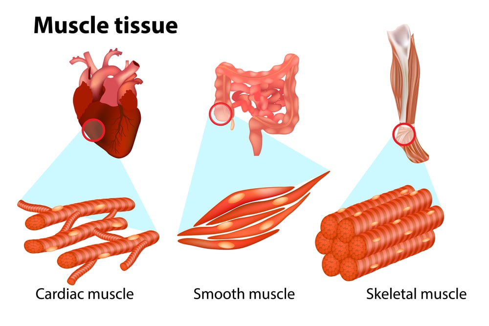
The cells of muscle tissue are elongated and often taper at their ends; hence these cells are called muscle fibres. The fibres are typically designed and constructed to produce a force of contraction. Thus, the tissue is contractile and able to produce movements. Based on structural and functional characteristics, the muscles are classified into three types:
- Striated or voluntary muscle;
- Smooth or involuntary muscle; and
- Cardiac muscle.
Striated Muscle
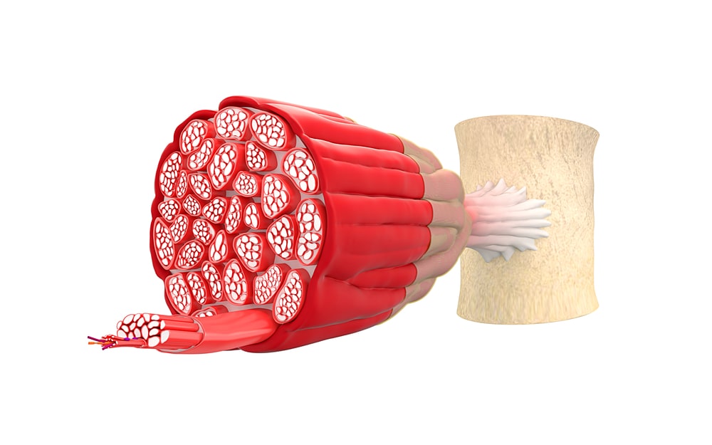
It is described as skeletal, striated, striped or voluntary muscle. It works under the control of the will. It is found in skeletal muscle. The cells of voluntary muscle, about 10-40 millimetres in length are roughly cylindrical. The sarcolemma is the fine sheath surrounding each muscle fibre and several nuclei are situated under it. The muscle fibres lie parallel to one another and show transverse dark and light bands.
The muscles consist of a large number of muscle fibres. Each fibre is enclosed in and attached to fibrous tissue called endomysium. Small bundles of fibres are enclosed in perimysium and the whole muscle in epimysium. The fibrous tissue extends beyond the muscle fibres to become the tendon which attaches the muscle to bone or skin.
Smooth Muscle
It is described as involuntary, plain or visceral muscle. It is not under the control of our will. It is made up of spindle-shaped cells, with only one central nucleus. There is no distinct sarcolemma but a very fine membrane surrounds each fibre. The bundles of cells form a sheet. During contraction and relaxation, the cells glide over one another. It is found in the walls of the blood vessel, lymph vessels, alimentary canal, respiratory tract, urinary bladder and uterus.
Cardiac Muscle
It is found exclusively in the wall of the heart. The cardiac muscle cells show cross stripes similar to those of voluntary muscle. Each cell has a nucleus and one or more branches. The ends of the cells and their branches are in close contact with each other forming “intercalated discs” which look like thicker and darker lines. This arrangement gives cardiac muscle an appearance of a sheet of muscle rather than fibres. Each fibre does not need to be stimulated as the impulse spreads from cell to cell across intercalated discs.
Make sure you also check our other amazing Article on: Epithelial Tissue