The peripheral nervous system consists of the following components:
- 31 pairs of spinal nerves
- 12 pairs of cranial nerves
- The autonomic part of the nervous system
Most of the nerves of the peripheral nervous system are composed of sensory nerve fibers conveying impulses from sensory organs to the brain. Motor nerve fibers convey impulses from the brain through the spinal cord to the effector organs like skeletal muscles, smooth muscles, and glands. Each nerve consists of numerous nerve fibers formed into bundles. Each bundle has several coverings of protective connective tissue.
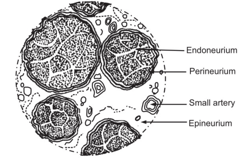
- Endoneurium is a delicate tissue surrounding each individual fiber and passes inwards from the perineurium.
- The perineurium is a smooth tissue, surrounding each bundle of fibers.
- Epineurium is the tissue that surrounds and encloses a number of bundles of nerve fibers. Most large nerves are covered by epineuria…
Table of Contents
Spinal Nerves
There are 31 pairs of spinal nerves, that leave from the vertebral column. The nerves pass through the intervertebral foramina formed by adjacent vertebrae. They are named and grouped as follows:
- 8 Cervical
- 12 Thoracic.
- 5 Lumbar
- 5 Sacral
- 1 Coccygeal.
Although there are only 07 cervical vertebrae, there are 08 cervical nerves; because the first pair leaves the vertebral canal between the occipital bone and the atlas and the eighth pair leaves below the last cervical vertebra. The lumbar, sacral and coccygeal nerves leave the spinal cords near its termination at the level of the first lumbar vertebra, and extend downwards inside the vertebral canal in the subarachnoid space, forming a sheath of nerves that resembles a horse’s tail, the caudal equine. These nerves leave the vertebral canal at the appropriate lumbar, sacral, or coccygeal level, depending on their destination.
Each spinal nerve is formed by the union of a motor and a sensory nerve root and is, therefore, termed a mixed nerve. Each spinal nerve is associated with the sympathetic part of the autonomic nervous system in the form of a preganglionic fiber.
Plexuses
Immediately after emerging from the intervertebral foramina, each spinal nerve divides into a ramus communicant, a posterior ramus, and an anterior ramus. The rami communicants are part of preganglionic sympathetic neurons of the autonomic nervous. systems. The posterior rami pass backward and divide into medial and lateral branches to supply skin and muscles of small areas of the posterior part of the head, neck, and trunk. The anterior rami supply the anterior and lateral parts of the neck, trunk, and upper and lower limbs.
In the cervical, lumbar and sacral regions, the anterior rami unite near their origins to form large masses of nerves or plexuses, where nerve fibers are regrouped and rearranged before proceeding to supply skin, bones, muscles, and joints of a particular area. In the thoracic region, the anterior rami do not form plexuses.
There are 05 large plexuses of mixed nerves formed on each side of the vertebral column. They are as follows:
- Cervical plexuses
- Brachial plexuses
- Lumbar plexuses
- Sacral plexuses
- Coccygeal plexuses.
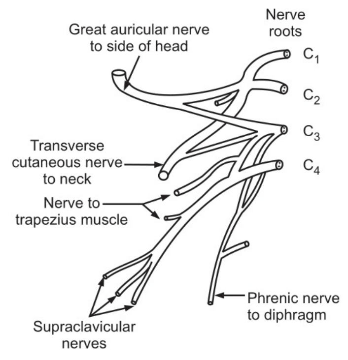
Cervical Plexus:
It is formed by the anterior rami of the first four cervical nerves. The superficial branches supply the structures at the back and side of the head and the skin of the front of the neck. to the level of the sternum. The deep branches supply muscles of the neck. The phrenic nerve originates from cervical roots 03, 04, and 05 and passes downwards through the thoracic cavity in front of the root of the lung to supply the muscles of the diaphragm.
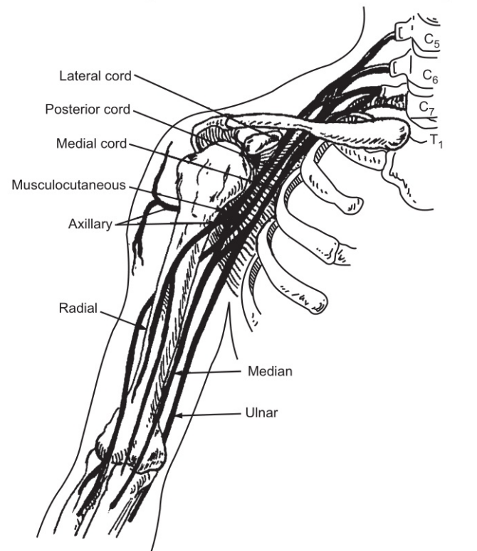
Brachial Plexus:
It is formed by the anterior rami of the lower four cervical nerves and a large part of the first thoracic nerve. It is situated above and behind the subclavian vessels and in the axillae. The branches of the brachial plexus supply the skin muscles of the upper limbs and some of the chest muscles. Five large nerves and a number of smaller ones emerge from this plexus. Each nerve has a contribution from more than one nerve root, containing sensory, motor, and autonomic fiber.
Following are the names of these nerves:
- Peripheral Nervous System
- Axillary nerve
- Radial nerve
- Musculocutaneous nerve
- Median nerve
- Ulnar nerve
- Medial cutaneous nerve
The axillary nerve supplies to the deltoid muscle, shoulder joint, and overlying skin. The radial nerve supplies the triceps muscle behind the humerus crosses in front of the elbow joint and further extends to the wrist and finger joints. It continues into the back of the hand to supply the skin of the thumb, the first two fingers, and the lateral half of the third finger. The musculocutaneous nerve supplies muscles of the upper arm and the skin of the forearm. The median nerve supplies the muscles of the front of the forearm. It continues into the hand where it supplies small muscles and the skin of the front of the thumb, the first two fingers, and the lateral half of the third finger. The ulnar nerve supplies the muscles on the ulnar aspect of the forearm. It continues to supply the muscles in the palm of the hand and the skin of the whole of the little finger and the medial half of the third finger.
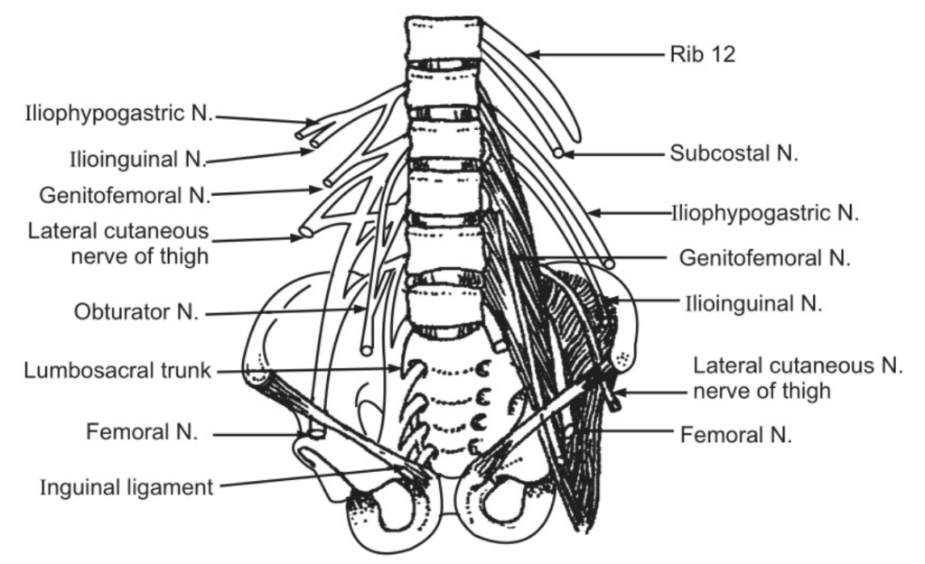
Lumbar Plexus
It is formed by the anterior rami of the first three and a part of the fourth lumbar nerve. The main branches and their nerve roots are:
- Iliohypogastric nerve
- Ilioinguinal nerve
- Genitofemoral nerve
- The lateral cutaneous nerve of thigh
- Femoral nerve
- Obturator nerve
- Lumbosacral trunk.
The Iliohypogastric, Ilioinguinal and Genitofemoral nerves supply impulses to the muscles and the skin in the area of the lower abdomen, upper and medial aspects of the thigh, and the inguinal region. The lateral cutaneous nerve of the thigh supplies impulses to the skin of the lateral aspect of the thigh including part of the anterior and posterior surfaces. The femoral nerve divides into cutaneous and muscular branches to supply the skin and muscles of the front of the thigh. One of the branches termed saphenous nerve supplies the medial aspect of the leg, ankle, and foot. The Obturator nerve supplies impulses to the adductor muscles of the thigh and skin of the medial aspect of the thigh. It aims above the level of the knee joints. The Lumbosacral trunk descends into the pelvis and supplies impulses to the sacral plexus.
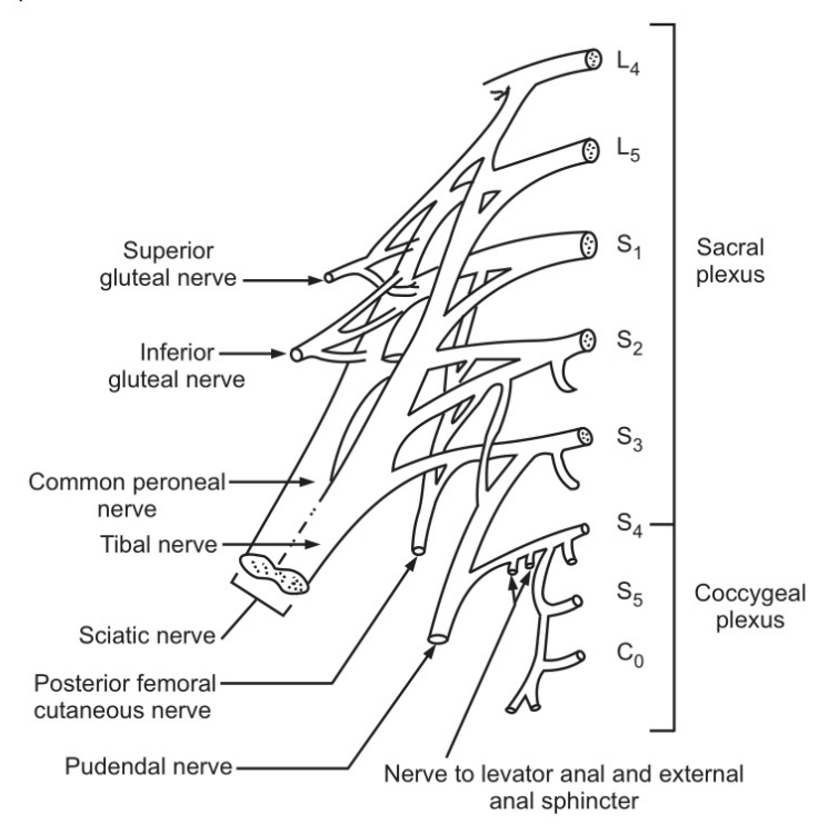
Sacral Plexus:
It is formed by the anterior rami of the lumbosacral trunk and the first, second, and third sacral nerves. The sacral plexus divides into a number of branches supplying impulses to the muscles and the skin of the pelvic floor, muscles around the hip joint, and the pelvic organs. It also provides the sciatic nerve.
The sciatic nerve supplies impulses to the hamstring muscles. At the level of the middle of the femur, it divides to form the tibial and the common peroneal nerves: The tibial nerve supplies impulses to the muscles and skin of the sole of the foot and toes. One of the main branches is the sural nerve which supplies the tissues in the area of the heel, the lateral aspect of the ankle, and a part of the foot. The common peroneal nerve after division into deep peroneal and superficial peroneal nerves, supplies impulses to the skin and muscles of the anterior aspect of the leg, the foot, and toes. The pudendal nerve supplies impulses to the external anal sphincter, the external urethral sphincter, and adjacent skin.
Coccygeal Plexus:
It is formed by a part of the 4th and 5th sacral and coccygeal nerves. It supplies impulses to the skin in the area of the coccyx and coccygeus muscles of the pelvic floor and the external anal sphincter.
Thoracic Nerves
These nerves do not mix to form plexuses. There are 12 pairs and the first 11 are the intercostal nerves, the 12th pair is a subcostal nerve.
Cranial Nerves
There are 12 pairs of cranial nerves originating from nuclei in the inferior surface of the brain. Some of them are sensory while others are motor in nature.
Autonomic Nervous System
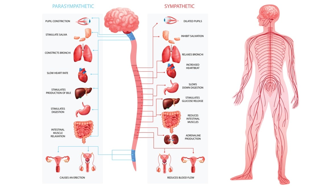
The autonomic nervous system primarily controls involuntary functions of the body which are carried out almost automatically. The stimulus for activities of the autonomic nervous system initiate in the brain below the level of the cerebrum. Although the actions are not voluntary in nature, the individual is aware of the actions exerted by the autonomic nervous system, e.g. variation in heart rate. The effects of the autonomic nervous system are essential for homeostasis and include stimulation or depression of glandular secretions and contraction of cardiac and smooth muscle tissues.
The efferent or motor nerves of the autonomic nervous system originate from nerve cells in the brain and emerge at various levels between the midbrain and the sacral region of the spinal cord. Many of them travel along the same nerve sheath, like that of the peripheral nerves of the central nervous system.
The autonomic nervous system is divided into the following two parts:
- Sympathetic nervous system (Thoracolumbar outflow)
- Parasympathetic nervous system (craniofacial outflow)
The two sub-parts have structural and functional differences. By and large, physiologically, they are opposite in functions; as a result, their working helps in maintaining. homeostasis and restoring the balance of the body. The sympathetic nervous system is said to be the part controlling ‘fight, flight, and fright’; while the parasympathetic nervous system controls activities related to maintenance and development during rest.
Sympathetic Nervous System
Three neurons are involved and convey impulses from their origin in the hypothalamus, reticular formation, and medulla oblongata to different effector organs and tissues. Neuron 01 has its cell body in the brain and its fiber extends into the spinal cords. Neuron 02 has its cell body in the lateral column of grey matter in the spinal cord, between the levels of the first thoracic and second or third lumbar vertebrae. Neuron 03 has its cell body in a ganglion and terminates in the organ or tissue.
Sympathetic Ganglia
Various sympathetic ganglia originate on either side of the spinal cord. These are chains of ganglia extending from the upper cervical level to the sacrum. The ganglia are attached to each other by nerve fibers. The fiber from the spinal cord up to ganglia is termed as preganglionic fiber, while the fiber from the ganglia to the effector organ is called postganglionic fiber.
Most of the organs are supplied with both sympathetic and parasympathetic fibers; however, there are few exceptions. Sweat glands, the skin, and blood vessels of skeletal muscles are n supplied with the parasympathetic nervous system. The effects of stimulation of various structures by the sympathetic nervous system and their consequent function are mentioned in Fig. 1.6.
There are three prevertebral ganglia situated in the abdominal cavity close to the arteries. Their names are as follows:
- Celiac ganglion
- Superior mesenteric ganglion
- Inferior mesenteric ganglion
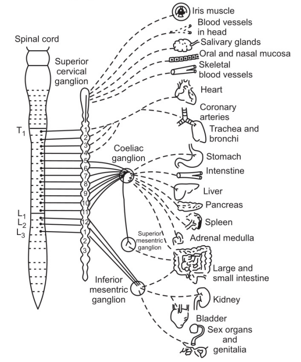
The ganglia consist of nerve cells diffusely distributed among a network of nerve fibers that form plexuses.
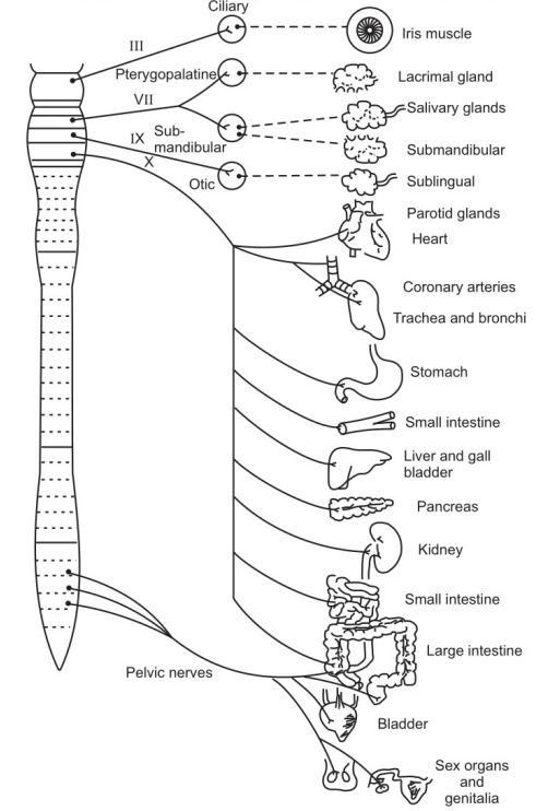
Parasympathetic Nervous System
Two neurons are involved in the transmission of impulses from their source to the effector organ. Neuron 01 has its cell body either in the brain or in the spinal cord. Those originating in the brain are the cranial nerves III, VII, IX, XI arising from nuclei in the midbrain and brainstem, and their nerve fibers terminate outside the brain. The cell body of the sacral outflow is in the lateral columns of grey matter at the distal end of this spinal cord. Neuron 02 has its cell body either in a ganglion or in the wall of the organ supplied.
Neurotransmitters
Noradrenaline is the chemical secreted at the postganglionic nerve endings of the sympathetic nervous system; the only exception to this is neuron 02 of the sympathetic nervous system. Acetylcholine is the chemical secreted at the postganglionic nerve endings of the parasympathetic nervous system. Both the chemical messengers of the autonomic nervous system are termed neurotransmitters. All ganglion transmissions, of the parasympathetic and sympathetic nervous systems, are mediated by acetylcholine.
Physiology of ANS
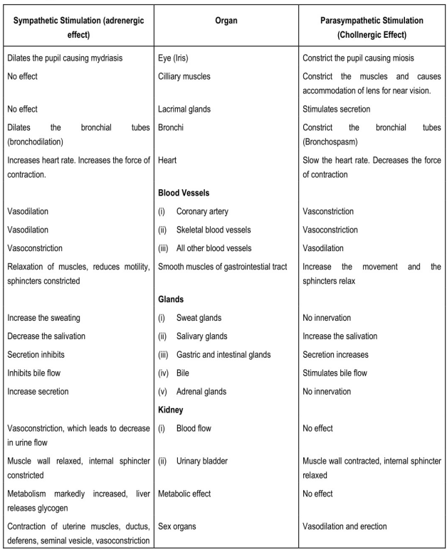
Note: Increased sweating and increased secretion of adrenal glands is cholinergic and not adrenergic though it is sympathetic stimulation.
Make sure you also check our other amazing Article on: The Joints