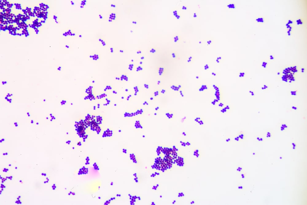
Simple staining is carried out to visualize bacteria and to compare morphological shapes and arrangements of the bacterial cell. In this technique, the bacterial smear is stained with a single basic dye such as crystal violet, safranin, methylene blue, etc.
Principle of the simple staining
These single basic dyes will give up a hydroxyl ion or accept a hydrogen ion that leaves the stain positively charged. Since the surface of bacteria is negatively charged so these charges are strongly bound to the cell surface (cell wall). Then the cells are visible against a light background.
Steps on the simple staining
- Clean and dry microscope slides thoroughly.
- Flame the surface on which the smear is to be spread.
- Flame the inoculating loop.
- Transfer a loop full of tap water to the flamed slide surface.
- Reflame the loop making sure the entire length of the wire that will enter the tube has been heated to redness.
- Remove the tube cap with the fingers of the hand holding the loop.
- Flame the tube mouth.
- Disperse the bacteria on the loop in the drop of water on the slide and spread the drop over an area the size of a dime. It should be a thin, even smear.
- Reflame the inoculating loop to redness including the entire length that entered the tube.
- Allow the smear to dry thoroughly.
- Fix the smear cautiously bypassing the underside of the slide through the burner flame two or three times.
- Stain the smear by flooding it with one of the staining solutions and allowing it to remain covered with the stain for the time designated below. Methylene blue – 1 minute Crystal violet – 30 seconds Carbol fuchsin – 20 seconds During the staining the slide may be placed on the rack or held in the fingers.
- At the end of the designated time rinse off the excess stain with gently running tap water. Rinse thoroughly.
- Wipe the back of the slide and blot the stained surface with a paper towel.
- Place the stained smear on the microscope stage smear side up and focus the smear using the 10X objective.
- Choose an area of the smear in which the cells are well spread in a monolayer. Center the area to be studied, apply the oil directly to the smear, and focus the smear under oil with the 100X objective.
Observation: Spherical-shaped bacteria coccus is turned blue color whereas rod-shaped bacteria, bacilli turn a red color.
Use: To study morphology and arrangement of bacteria cells.
Make sure you also check our other amazing Article on : Methods of isolation of pure culture