Bacteria are microscopic, single-celled organisms that exist in their millions, in every environment, both inside and outside other organisms. Some bacteria are harmful but must serve a useful purpose. They support many forms of life, both plant, and animal, and they are used in industrial and medicinal processes.
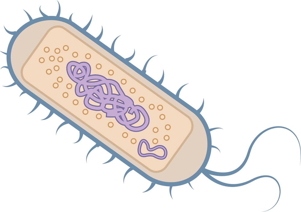
Under the electron microscope, the structure of the bacterial cell looks like a capsule. It has the following components like outside the cell membrane and internal to the cell membrane (Fig. 1)
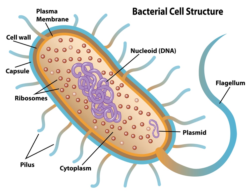
Table of Contents
Capsule:
It is the outer layer of the bacteria cell. Depending on the chemical nature capsules are thick or thin, and rigid or flexible. It is the most important virulence factor of bacteria which is visualized by the negative staining technique. Generally, it is composed of 98% water and 2% polysaccharide or glycoprotein/polypeptide or both. Only some bacterial species possess capsules. The capsule is usually made of polysaccharides (e.g. pneumococcus), occasionally polypeptide (e.g. anthrax bacilli), and hyaluronic acid (e.g. streptococcus). Most bacterial capsules are composed of polysaccharides especially homopolysaccharides which are usually synthesized outside the cell from disaccharides by the exocellular enzyme. The capsule of acetic acid bacteria cell is composed of homopolysaccharide (hemicellulose).
Glucan is synthesized by Streptococcus mutants which are composed of several types of sugar and are termed heteropolysaccharides. A few capsules are polypeptides like Bacillus anthracis, which is composed entirely of a polymer of glutamic acids.
There are two types of capsules
- Microcapsule: Thickness of 0.2 µm or more, visible under a light microscope.
- Microcapsule: Thickness is less than 0.2 µm, visible under Electron microscope.
Functions:
- Preventive nature: Capsular polysaccharide binds a significant amount of water making cells resistant to drying and also preventing attachment of bacteriophage on the cell surface.
- Protection: It protects from mechanical injury, temperature, drying, etc.
- Attachment: Capsule helps in attachment on the surface. For example, Streptococcus mutants that cause dental carries attach on teeth surface by its capsule.
- Anti-phagocytic: Capsule resists phagocytosis by WBCs.
- Source of nutrition: It acts as a resource organ of nutrition during insufficient nutrient supply.
- Repulsion: Same charge capsulated bacteria repel each other.
Cell wall:
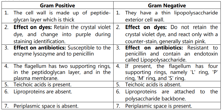
The cell wall is the rigid wall situated outside of the plasma membrane. It provides the shape of the bacteria as well as protects from osmotic lysis. The cell wall is made up of peptidoglycan. The gram-positive bacteria contain thick homogeneous peptidoglycan or murein layer (20 to 80 mm thickness) outside the plasma membrane whereas gram-negative bacteria have a 2-7 nm peptidoglycan layer surrounded by a 7-8 nm thick outer membrane. Peptidoglycan is a polymer consist of two sugar molecules viz. N-acetylglucosamine and N-acetylmuramic acid. Apart from that it also contains several amino acids.
Flagella:
Flagella are slender and rigid hair-like structures. They are about 20 nm across and up to 15 to 20 µm long. The eukaryotic flagellum is a long, rod-like structure, surrounded by an extension of the cell membrane like a sheath. The bulk of the structure is a filament called an axoneme. Monotrichous bacteria have one polar flagellum whereas amphitrichous bacteria have either singe or duster of flagellum at both poles. Lophotrichous bacteria have a cluster of flagella at one end. Peritrichou bacteria have surrounded lateral flagella.
Flagellar Ultra-Structure:
Transmission electron microscope studies revealed three regions of flagella which are:
- The outermost region is the filament which is extended from the cell surface to the tips.
- Basal bodies consist of small central rods inserted into cells
- A short curved segment, the flagellar hook which links the filament to basal bodies and acts as flexible coupling.
The filament is a hollow, rigid cylinder made up of protein subunits flagellin. The molecular weight varies from 30,000 daltons to 6,000 daltons depending on the bacterial species.
Gram-negative bacteria have four rings connected to a central rod. Two rings (M and S rings) are embedded in the cell membrane and the other two rings (L and P rings) are associated with the cell wall which is made up of lipopolysaccharide and peptidoglycan respectively. Gram-positive bacteria have only two rings in their basal body. M-ring which is an inner ring, connected to the plasma membrane, and S-ring which is the outer ring, connected to the peptidoglycan.
Fimbriae and Pili:
Many Gram-negative bacilli contain short, fine, hair-like non-flagellar appendages called fimbriae. Pili are similar appendages which are about 1-10 cells. The filament of pilus is straight and the diameter is 7 nm to 10 nm, larger than the diameter of fimbriae. It is made up of pilin protein. Molecular weight is 17,000. Pili is a non-motile but adhesive structure. They enable the bacteria to stick firmly to other bacteria, to a surface, or some eukaryotic such as mold plants, plants, and animal cells. They are genetically determined by sex factor or conjugative plasmid and hence they help in the conjugation of male bacteria, in the attachment of pathogenic bacteria to their host cell.
Spinae:
Some Gram-positive bacteria have tubular unicellular and rigid appendages of a single protein moiety, known as spinae. They help the bacterial cell to tolerate environmental stress such as salinity, pH and temperature, etc.
Cell Membrane:
Cell or plasma membrane is a thin structure that surrounds the cell. It is about 8 nm thick. This structure is a critical barrier separating the inside of the cell from the environment. The cell membrane is also a highly selective barrier enabling the cell to concentrate a specific metabolite and excrete waste material. Mostly biological membrane is composed primarily of Phospholipids (about 20 to 30 percent) and proteins (about 60 to 70 percent).
One major difference in the chemical composition of membrane between eukaryotic cells and prokaryotic cells is that eukaryotes have sterol in their membrane depending on cell type, sterol can make up from 5-25% of the total lipid of the eukaryotic membrane. Sterols are absent from membranes of all prokaryotic cells (methanotrophs are a major exception). Sterols are rigid, planner molecules, whereas fatty acids are flexible.
The main function of the cell membrane is selective permeability and transport of solute, electron transport and oxidative phosphorylation in aerobic species, excretion of hydrolytic exoenzymes.
Inclusion bodies:
Various types of inclusion bodies organic and inorganic granules are present in the cytoplasmic matrix. They are mainly used for the storage of energy and reduce osmotic pressure by tying up molecules in particulate forms like polyphosphate granules, glycogen granules, etc. Some of the membrane-enclosed inclusion bodies are glycogen-sulfur granules, carboxysomes, etc.
In many cyanobacteria, green photosynthesis bacteria gas vacuole is present as an organic inclusion body. Gas vacuoles are aggregates of a huge number of small, hollow cylindrical structures and provide them buoyancy.
Ribosomes:
Ribosomes are loosely attached to the plasma membrane (70S ribosomes). They are made up of both protein and ribonucleic acid. They are the site of protein synthesis. Plasma membrane ribosomes make proteins for transport to the outside.
Nucleoid:
A prokaryotic chromosome is located in an irregularly shaped region called the nucleoid. Nucleoid is composed of 60% DNA, 30% RNA, and 10% protein by weight. Prokaryotes contain a single circle of double-stranded DNA but some have linear DNA chromosomes.
Mesosome:
Mesosomes are present in the cell membrane of bacterial cells that fold inward. They play a major role in cellular respiration. They appear as a pocket formed by the cytoplasmic membrane, filled with vesicles, tubules, or lamellae.
Some of the differences between gram-positive and gram-negative bacteria are given in Table…
Make sure you also check our other amazing Article on : Classification of Microorganisms