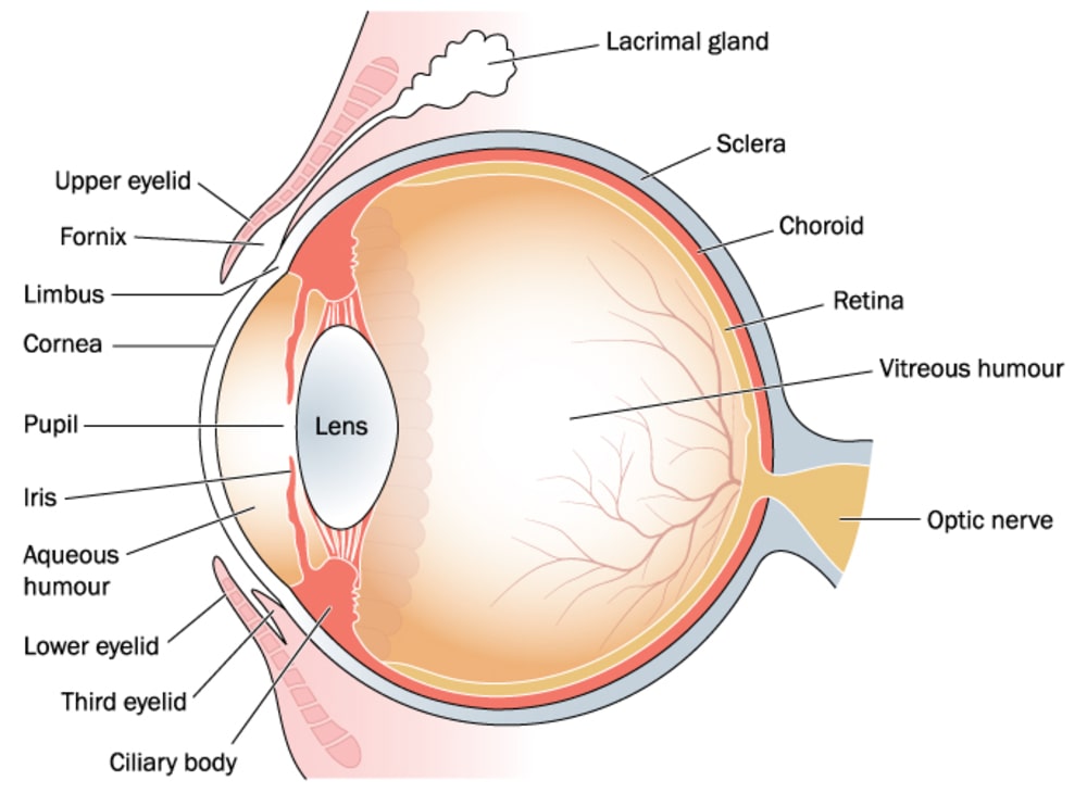
Anatomy of Eye is a special sense organ of sight present in the orbital cavity. It is spherical in shape and about 2.5 cm in diameter. The orbit of the eye is formed by different bones which protect the eyeball.
Anatomically, the wall of the eyeball is divided into three layers.
- Outermost layer is the fibrous coat, consisting of sclera and cornea.
- The middle layer is a vascular coat, consisting of the choroid, ciliary body, and iris.
- The innermost layer is made up of nervous tissue, consisting of the retina.
There are six muscles, called as external muscles. These muscles connect the eyeball to the orbital cavity. These six muscles are four rectus muscles, i.e. superior, inferior, lateral and medial and two oblique muscles, superior and inferior.
Anterior to the eyeball is eyelids, which protect the eyeball from dust and intense light. At the margin of the eyelid, eyelashes are present which further protect the eyeball. The mucous membrane which lies in the inner surface of each eyelid is known as the conjunctiva. This conjunctiva covers the exposed area of the eyeball.
Table of Contents
Eye Structure
The Lachrymal Apparatus
It is composed of the lachrymal gland, where tears are produced. The lacrimal canaliculi, lachrymal sac and nasal lachrymal ducts, all assist in carrying tears to the nasal cavity. The lachrymal glands are present in the depression of the frontal bone. Each gland is provided by about twelve ducts. These ducts open at the surface of the conjunctiva at the upper-eyelid where fluid has pored. This fluid has an anti-bacterial action that protects the eye from foreign materials.
The Sclera and Cornea
The sclera is the outermost coat of the eye consisting of fibrous tissue. This layer is tough and maintains the shape of an eye. It also gives attachment to the extrinsic muscle of the eye. But anteriorly, the sclera becomes a transparent membrane called as the cornea, made of epithelial tissue. This part of the cornea is used for passing light rays to reach at the retina. The cornea is convex anteriorly, used for bending the light rays to focus them on the retina.
The Choroid
The choroid is the middle coat which lines the sclera inside the posterior for about five-sixths part. It is a very highly vascularized part, deep chocolate brown in colour. The light rays pass into the eye through the pupil and the nerve endings in the retina are stimulated by light rays. The choroid provides nutrients to the posterior part of the retina. The chocolate brown colour of the choroid is mainly due to pigment, melanin which is produced by melanocytes.
The anterior part of the choroid contains the ciliary body which consists of non-striated muscle fibres, i.e. ciliary muscles and also contains epithelial cells. The ciliary body contains ciliary processes, which is an extension of the internal surface of the ciliary body, containing blood vessels. The epithelial cells secrete a watery fluid known as aqueous humour. The circular band of smooth muscle is called ciliary muscle which helps in altering the shape of the lens for far or near vision. The aqueous humour is present in front of the lens. The nerve supply to the ciliary body is from the parasympathetic nerve, the branches of the Oculomotor nerve.
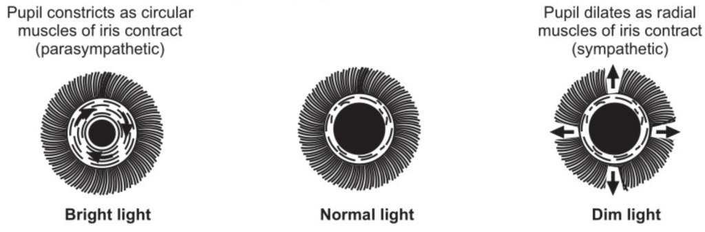
The iris commences anteriorly from the ciliary body which covers the anterior one-sixth of the eye. Iris is present between cornea and lens and is attached to the outer margin of ciliary processes. Iris consists of circular and radial smooth muscles. The aperture in the centre of the iris is called as a pupil.
The main function of the iris is to control the light rays entering in the eye. When there is a bright light, the parasympathetic neuron stimulates the circular muscle of the iris to contract which helps to decrease the size of the pupil whereas in dim light, the sympathetic neuron stimulates the radial muscles of the iris to contract to cause dilating the pupil. The dilation and constriction of the pupil are autonomic reflexes. The lens is a circular – biconvex transparent body. The suspensory ligaments from the ciliary body hold the lens and is enclosed within the capsule. The lens is elastic in nature and lies behind the pupil. The thickness of the lens is controlled by the ciliary muscle through the suspensory ligament.
The Retina
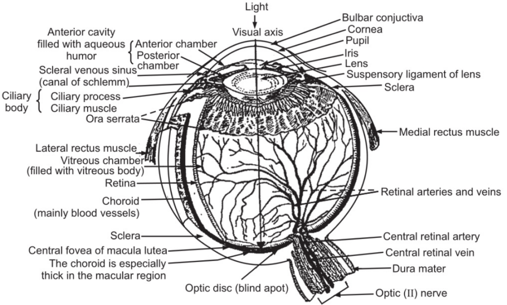
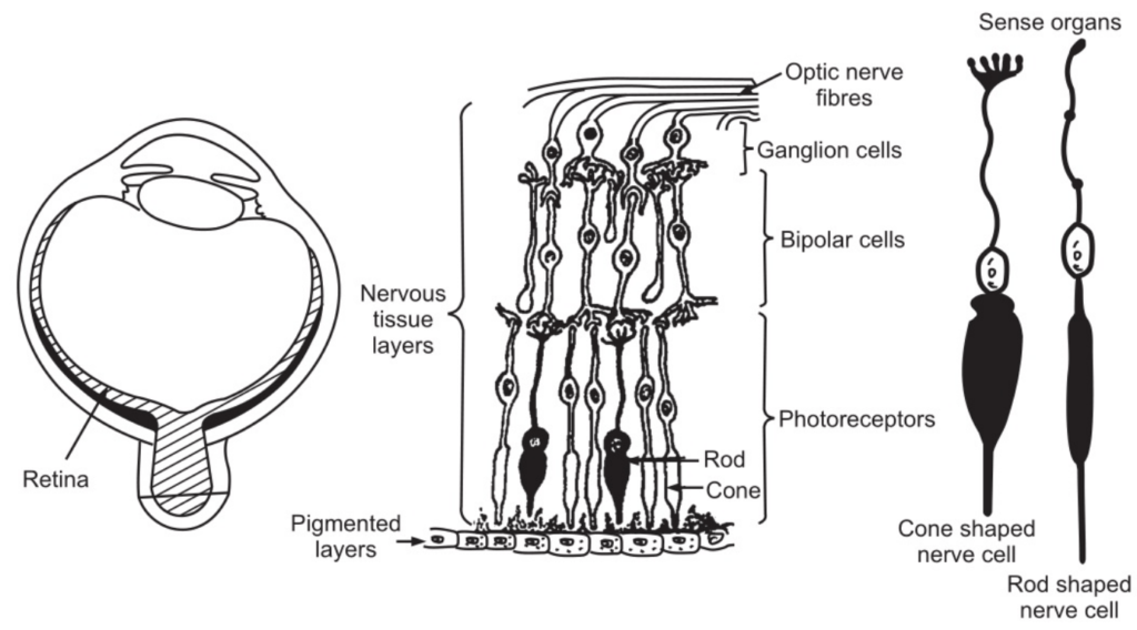
The retina lines the inner surface of the choroid and is very delicate and easily stimulated by light rays. The retina is composed of nerve cells and nerve fibres and these nerve cells and nerve fibres are mounted on pigmented layers of epithelial cells and are attached to the choroid. Nerve cells and nerve fibres are called rods and cones that are highly sensitive to light rays. A magnified image of the retina can be seen through the pupil by an ophthalmoscope. During hypertension and diabetes, some pathological changes in blood can be observed. At the posterior end inside of the eyeball, the retina occupies three-quarters of space. A very thick layer is present at the back and it thins out anteriorly up to the ciliary body. At the centre of the retina, there is a small depression containing yellow coloured cells known as macula lutea. Fovea centralis is present at the centre, having a small depression which contains only cone-shaped cells. This portion is a very sensitive part of the retina. As the retina advances to the anterior side, there are more rod-shaped cells and very few cone-shaped cells. Retina has slight purple colour due to the presence of visual purple in the rods. Near the macula lutea, the fibres of the retina come together to form the optic nerve which passes out backside to the eye-ball through the sphenoid bone to reach the occipital lobe of the cerebrum. Here at the optical nerve where it leaves the eye, light-sensitive cells are absent, this area is called an optic disc or blind spot.
The ciliary arteries and central arteries supply the oxygenated blood to the eye and they come from the ophthalmic artery which is also a branch of the internal carotid artery. The central retinal vein drains venous blood from the retina through the optic disc. Retina has pigment epithelium and neural part. Melanin is present in the choroid and pigment epithelium in the retina; absorbs light rays, and helps to prevent scattering of light within the eyeball. This ensures that the image produced on the retina by the cornea and lens remains sharp and clear. The neural part of the retina is the multilayered outgrowth of the brain. The retina consists of about 06 million cones and 120 million rods. Rods help in observing shades of grey in dim light and also help to see shape and movement. Cones help in colour vision in bright light. Colours cannot be seen in the moonlight as only rods are functioning because the light is too less to stimulate the cone.
At the anterior side of the eye, there is a space between the cornea and lens. This part is completely divided into anterior and posterior chambers by the iris. Both these chambers are filled up with watery fluid called aqueous humour. At the back of the lens, the eyeball cavity is filled with vitreous humour which is soft, colourless, and transparent and a jelly-like substance; contains 99 per cent water, some salts and protein. Vitreous humour helps in maintaining intraocular pressure to support the retina against the choroid.
Rods
Rods are very sensitive to dim light and their function is in night vision. Rod consists of a photosensitive chemical known as rhodopsin. Rhodopsin is a combination of vitamin ‘A’ (retinol) and a protein called pepsin. But when the retina is exposed to bright light rhodopsin vanishes. Regeneration of rhodopsin occurs under reduced illumination which helps in the adjustment of the eyes to dim light, known as dark adaptation. During this process, the iris increases the diameter of the pupil. Vitamin A deficiency shows the shortage of rhodopsin which causes night blindness, i.e. inability to see objects in the night (dim light).
Cones
Cones are essential for daylight vision and colour vision. There are three types of cones, having different pigments, most sensitive to red, blue or green light. When there is stimulation in correct proportion in the three types of cones, the sensation is white. When one type of cone is stimulated, there is the sensation of the light, corresponding to the type of the cone stimulated. When two types of cones are stimulated, the sensation is of a colour. Normal colour vision mainly depends upon the combination of these three primary colours in the sensory cortex. In colour blindness, the individual cannot differentiate between red and green because there is a deficiency of red-sensitive or green-sensitive pigment.
Physiology of Vision
When an image of the object is focused on the retina, it stimulates the photoreceptors which transfer the light stimulus onto the bipolar cells. The bipolar cells communicate with ganglion cells which extend their axons to the lateral geniculation body of the thalamus. Fibres carrying visual nerve impulse from the thalamus goes to the primary visual cortex in the occipital lobe.
The outer part of segments of cones is cone-shaped whereas those of rods are rod-shaped. The conversion of light into an electrical signal occurs in the outer part of the segment. The nucleus is present in the inner part of the segment. Golgi complex and mitochondria are also present. The photoreceptor is expanded into a bulk-like synaptic terminal at the proximal end and the light is absorbed by the photopigment. These are coloured proteins and are present in the outer segment membranes. These proteins, due to absorption of light, undergo structural changes and the changes help for the production of the receptor potential. There is photopigment present in the rods is known as rhodopsin. The photo proteins are present in the plasma membrane. Some of the photo proteins form discs from the plasma membrane. Normally, 1000 discs are present in the outer segments of each rod. The photoreceptor at the outer segments is renewed at an astonishingly rapid pace. Every hour about one to three discs are added at the base of the outer segments and old discs are cast out at the tip and phagocytosed by pigment epithelial cells. The photoprotein contains a glycoprotein called opsin and a derivative of vitamin A known as retinal. The individual can see better by taking an adequate diet such as carrots, spinach or meats etc. which contain vitamin A. Deficiency of vitamin-A can cause night blindness. The visual photopigment is responsible for absorbing the light.
Optic nerve track
The optic nerve fibres originate from the retina of the eye and converge at a point approximately 0.5 cm to the nasal side of the macula lutea to become the optic nerve. The optic nerve passes through the choroid and sclera and goes backwards and medially through the orbital cavity, and then it goes backwards through the optic foramen of the sphenoid bone and centrally to meet the nerve from the other eye at the optic chiasm. The optic chiasm is present in front and above the pituitary gland. The optic nerve fibres in the optic chiasma come from the nasal side of each retina, cross over to the opposite side. Those fibres coming from the temporal side of the retina do not cross but continue backwards on the same side.
Optic tract
It is the pathway at the backside of the optic chiasma. The tract consists of nasal fibres from the retina of one eye and temporal fibres of the retina of other eyes. Tracts go backwards to meet a group of nerve cells called lateral geniculation bodies in the cerebrum. Lateral geniculation bodies lie behind and below the thalamus which works as a relay station for the optic nerve. The nerve fibres work as optic radiations and go backwards and divide in the visual area of the cerebral cortex in the occipital lobes of the cerebrum.
Accommodation
When the surfaces of the lens are curved outward (convex) then the lens refracts incoming rays towards each other so that they can intersect. But when the surface of the lens is curved inward, then the rays bend away from each other. This position of the lens is concave. The focusing power depends upon the increase or decrease in curvature of the lens. When a person wants to focus on near objects, the lens curvature increases to bend the rays towards the fovea centralis. This increase in the curvature of the lens, to see near objects is known as accommodation. When the eyes have to focus on distant objects, the ciliary muscle relaxes and the lens becomes flatter because it is stretched in all directions by suspensory ligaments. To focus on near objects, the ciliary muscles contract and pull the ciliary processes and choroid forward towards the lens. This releases the tension in the lens and suspensory ligaments. All these changes can occur due to the elastic nature of the lens.
When the light rays from an object enter the eyes, they go in at a different angle. But it is important that the light rays should stimulate two retinae. If this does not occur, then a person complains about double vision. When a person sees a near object, both the eyeballs move inwards so that their visual axes converge on the object. So nearer the object, the greater is the convergence. But when a person sees a distant object, less convergence is necessary. All these movements of the eyeball occur due to extrinsic muscles of the eyes.
Eye Abnormalities
Myopia or Nearsightedness
When there is a long distance between the cornea and lens or lens and retina or sometimes there is too powerful a lens, the person suffers from myopia. In this particular situation, the image of a distant object falls in front of the retina. That is, when the object distance is 20 feet, light rays go into the eye almost parallel. In such type of condition, an individual has to wear a concave lens to adjust the light rays coming from a distant object, so that image can fall on the retina.
Hyperopia or Farsightedness
When there is a short distance between cornea and lens or lens and retina or there is a weak lens, the person suffers from Hyperopia. The image of a distant object falls behind the retina. In such situations, by using a proper convex lens the image of the object can fall on the retina. Presbyopia
The old persons suffer from such a condition. In this, the lens slowly loses its elasticity and interferes with correct accommodation. By using the convex lens, accommodation can be corrected.
Cataract
As a person’s age advances, this condition interferes with vision. There is lens opacity and loss of transparency due to the scattering of light in this region. It may be due to certain disease conditions or drugs or exposure to X-rays.
Glaucoma
When there is excessive accumulation of aqueous humour, the intraocular tension increases; this can lead to severe eye disease, the retina may get damaged and the person suffers from blindness.
Conjunctivitis
Conjunctivitis may occur due to bacteria or dust, this may lead to inflammation and an increase in tear flow.
Colour blindness
This is a sex-linked inherited disease. About 0.4 per cent of the female population and about 08 per cent of the male population suffer from this abnormality. Some of the people are totally colourblind and every object is seen in terms of black and white and some are partially colourblind and can sense only some colours.
Night Blindness
It is caused due to deficiency of vitamin A. Such a person cannot see in dim light.
Make sure you also check our other amazing Article on: Lymph Nodes