Lymph nodes are present at strategic positions throughout the body. The small lymph vessels carry lymph towards these nodes. From here, the lymph passes into the blood.
Table of Contents
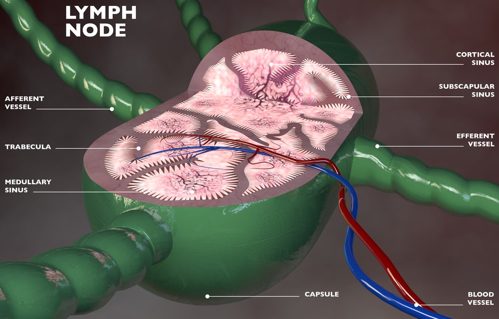
Structure of Lymph Nodes
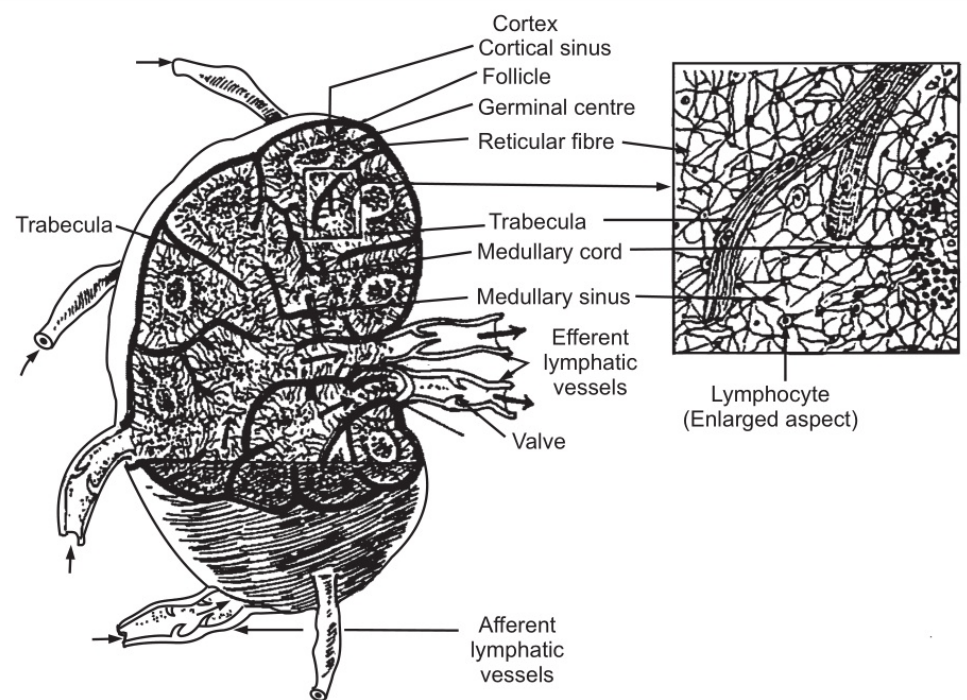
These are oval or bean-shaped organs present along the length of the lymphatic vessels. The size of the lymphatic gland is variable and ranges from 01-25 mm in length. These glands are found in maximum numbers in the axillae, groins, and mammary glands.
The node has a coating of dense connective tissue and these tissues extend into the node. These extensions are known as trabeculae. The trabeculae help to divide the nodes into different compartments, provide a passageway to blood vessels and give support. The capsule, trabeculae, reticular fibers, and fibroblasts, form the framework of the lymph node. The lymph node has two parts: the outer part is the cortex and the inner is the medulla. The cortex consists of many follicles that serve as a site for densely packed lymphocytes. The outer rim of each follicle contains T-cells i.e. T-lymphocytes and macrophages. The central area of the follicles is lighter where B-lymphocytes proliferate into antibody-secreting plasma cells. In the medullar region, the lymphocytes are tightly packed in strands known as medullary cords. These cords also consist of plasma cells and macrophages. The afferent lymph vessels enter the lymph node and efferent vessels carry lymph from the node. Being a bean-shaped organ, it has a concave surface, called the hilum through which the arteries pass and veins and efferent lymph vessels leave.
Functions of lymph nodes
During the passage of the lymph through the lymph node, the lymph is filtered. Some of the particulate matter containing microbes, dead and live phagocytes containing ingested micro-organisms, broken or damaged tissues, inhaled particles, etc. are filtered. Those particles which are not filtered enter the blood. Incomplete phagocytosis of microbes causes inflammation and enlargement of the lymph nodes. The multiplication of T and B lymphocytes occurs in the lymph node.
The Spleen
The spleen is the largest lymph organ and it is formed by lymphatic and reticular tissues. The spleen is located in the left hypochondriac region of the abdominal cavity. It lies between the diaphragm and fundus of the stomach. The average weight of the spleen is 200 gm. It is 12 cm long, 07 cm in width, and 02.5 cm thick.
Structure of the spleen
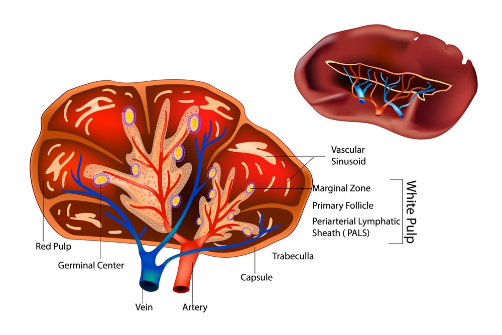
It is oval in shape. The hilum lies on the lower medial broader. The spleen is covered with fibroelastic tissue called as a capsule. The fibroelastic tissue extends inwards to form trabeculae. The splenic pulp is composed of lymphocytes and macrophages. This lies between the trabeculae. The pulp has a red portion (red pulp) and a white portion (white) pulp) which consists of lymphatic tissues. Lymphocytes and macrophages are seen in this part. The splenic artery and nerves enter the spleen while the splenic vein and lymph vessels (efferent vessels) leave the spleen. The splenic pulp is supplied by blood through the sinuses which have pores in endothelial cells.
Functions of Spleen
The old erythrocytes or abnormal red blood cells are destroyed by the spleen. Hemoglobin from the destroyed erythrocytes gets separated into various constituents such as bilirubin, biliverdin, and iron. These components are carried towards the liver via the portal veins Other components such as micro-organisms platelets, leucocytes are destroyed or phagocytosed by the spleen. In the fetal stage, the spleen produces erythrocytes.
Make sure you also check our other amazing Article on: Blood Groups