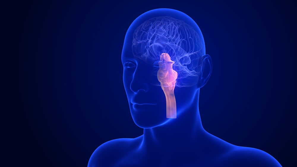
The posterior stalk-like region of the brain that connects the cerebrum to the spinal cord is known as the brainstem (or brain stem). The brainstem is made up of the midbrain, pons, and medulla oblongata in the human brain. Through the tentorial notch, the midbrain connects to the thalamus of the diencephalon, and the diencephalon is sometimes included in the brainstem.
Table of Contents
Midbrain
It is situated around the cerebral aqueduct between the cerebrum above and the Pons varolli below. It consists of groups of nerve cells and nerve fibres. It connects the cerebrum with the lower parts of the brain and the spinal cord. The nerve cells act as relay stations for the ascending and descending nerve fibres.
Pons varolli
It is situated in front of the cerebellum, below the midbrain and above the medulla oblongata. It consists of nerve fibres forming a bridge between the two hemispheres of the cerebellum, and of fibres passing between the higher levels of the brain and the spinal cord. Few groups of cells within the Pons act as relay stations. Some of them are associated with the cranial nerves. The nerve cells in the Pons lie deep and the nerve fibres are on the surface.
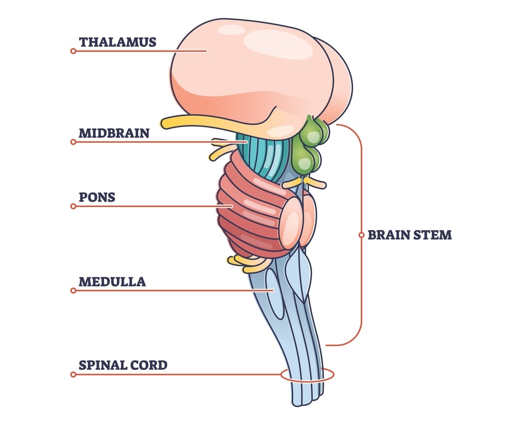
Medulla oblongata
It extends from the Pons varolli above and is continuous with the spinal cord below. It is shaped like a pyramid with its base upwards and it lies within the cranium above the foramen magnum. Its anterior and posterior surfaces are marked by central fissures. Some cells of the medulla oblongata constitute relay stations for sensory nerves passing from the spinal cord to the cerebrum. Following vital centres which are associated with autonomic reflex activity lie in its deeper structure.
- Cardiac centre
- Respiratory centre
- Vasomotor centre
- Reflex centres of vomiting, coughing, sneezing and swallowing
Medulla oblongata has the following special features:
Decussating of the pyramids: Motor nerves descending from the motor area in the cerebrum to the spinal cord in the pyramidal or corticospinal tracts, cross from left side to right and vice versa. These tracts are the main pathways for impulses to voluntary, i.e. skeletal muscles.
Sensory decussating: Some of the sensory nerves ascending to the cerebrum from the spinal cord cross from the left side to the right and vice-versa in the medulla oblongata. Few other sensory nerves decussate at the level of the spinal cord.
The cardiac centre: It controls the rate and force of cardiac contraction. Sympathetic and parasympathetic nerve fibres starting from the medulla pass to the heart.
The respiratory centre: It controls the rate and depth of respiration. From this centre, nerve impulses pass to the Phrenic and intercostal nerves which stimulate contraction of the diaphragm and intercostal muscles, thus initiating inspiration. The centre is stimulated by excess CO2 or by deficiency of O2 in the blood supply. Variation in concentrations of CO2/02 is conveyed by nerve impulses from the chemoreceptors in the carotid bodies.
The vasomotor centre: It controls the diameter of the blood vessels. It has special control over small arteries and arterioles which influence a large proportion of smooth muscle fibres in their walls, e.g. the sources of stimulation of the vasomotor centre, the arterial baroreceptors, body temperature and emotions such as sexual excitement and anger. The pain usually causes vasoconstriction; however, severe pain may cause vasodilatation, a fall in blood pressure and fainting.
Reflex centres: When irritating substances are present in the stomach or respiratory tract nerve impulses pass to the medulla oblongata, stimulating the reflex centres; this, in turn, initiates reflex actions of vomiting, coughing and sneezing.
Reticular Formation
It is a collection of neurons in the core of the brainstem, surrounded by neural pathways which pass nerve impulses between the brain and the spinal cord. It constantly receives information from the brain and the spinal cord and transmits it in descending and ascending tracts. It has the following functions:
- Co-ordination of skeletal muscle activity is associated with voluntary motor movement and the maintenance of balance.
- Co-ordination of activity control by the Autonomic Nervous system, e.g. cardiovascular, respiratory and gastrointestinal activity.
- Selective awareness functions through the Reticular Activating System (RAS). This can selectively block or pass sensory information to the cerebral cortex.
Make sure you also check our other amazing Article on: Anatomy of Brain