The brain lies within the cranial cavity. It constitutes about 1/5th of the body weight. It is composed of the following parts:
- Cerebrum or forebrain
- The brainstem
- Midbrain
- Pons varolli
- Medulla oblongata
- Cerebellum or hindbrain
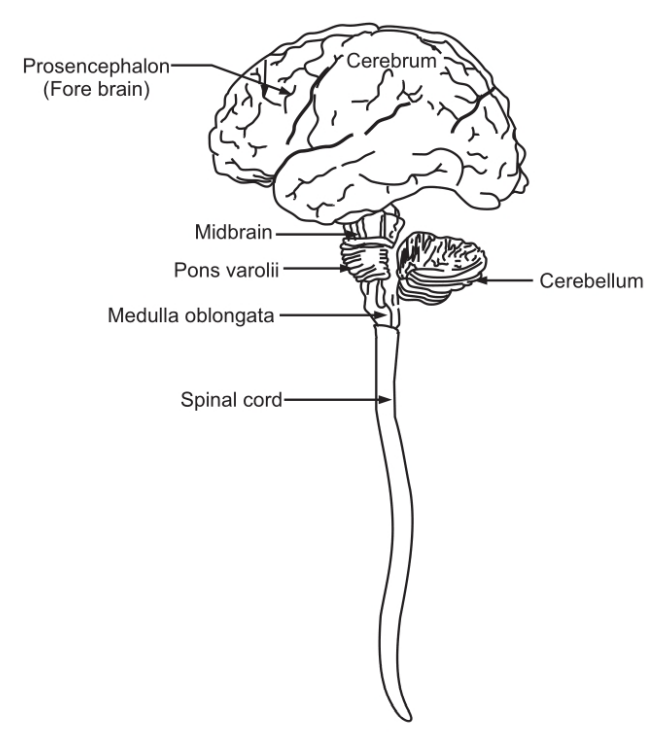
Table of Contents
Cerebrum
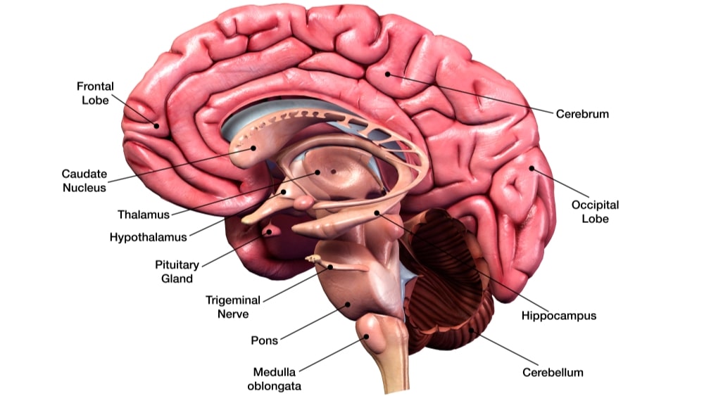
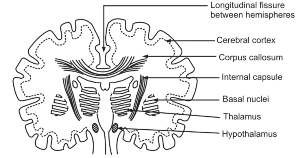
It is the largest part of the brain and occupies the anterior and middle part of the cranial cavity. It is divided by a deep cleft into the right and left cerebral hemispheres. Deep within the brain, the hemispheres are connected by a mass of nerve fibers called the corpus callosum. The superficial part of the cerebrum is composed of the nerve cell body, forming the cerebral cortex.
The cerebral cortex shows many enfolding of varying depth. The exposed areas of the fold are called as gyro or convolutions and are separated by sulci or fissure. The convolutions help in increasing the surface area of the cerebrum. Each hemisphere of the cerebrum is divided into the following lobes:
- Frontal
- Parietal
- Temporal
- Occipital
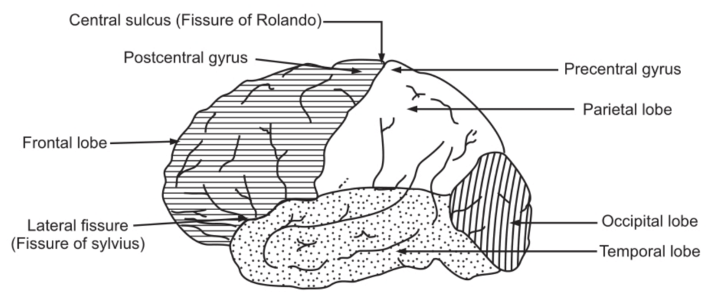
The boundaries of the lobes are marked by deep sulci; called as central, lateral, and parietal occipital sulci.
Within the cerebrum, the lobes are connected by masses of nerve fibers, or tracks, which constitute the white matter of the brain. Various afferent (incoming) and efferent (outgoing) fibers linking the different parts of the brain and spinal cord are as follows:
- Arcuate (association) fibers: These fibers connect different parts of a cerebral hemisphere by extending from one gyro to another.
- Commissural fibers: These fibers connect corresponding areas of the two cerebral hemispheres. The largest commissural is called as Corpus callosum.
- Projection fibers: These fibers connect the cerebral cortex with grey matter of lower parts of the brain and with the spinal cord. The internal capsule is one of the projection fibers. It lies deep within the brain between the basal ganglia and the thalamus. All nerve impulses passing to and from the cerebral cortex are carried by fibers that form the internal capsule. Motor fibers within the internal capsule form the pyramidal tracts (corticospinal tracts) that decussate at the medulla oblongata…
Functions of the Cerebrum
- Mental activities involved in memory, intelligence, sense of responsibility; thinking, reasoning, moral sense, and learning…
- Sensory perception, including the perception of pain, temperature, touch; sight, hearing, taste, and smell.
- Initiation and control of voluntary muscle contraction.
Functional Areas of the Cerebrum
Functional areas of the cerebrum are divided into sensory and motor areas. Anatomically there is a mixture of both these functions.
Motor Areas
The precentral area lies in the frontal lobe immediately anterior to the central sulcus. The nerve cells are pyramid-shaped and they initiate the contraction of voluntary muscles. A nerve fiber from a pyramid-shaped cell passes downwards through the internal capsule to the medulla oblongata where it crosses to the opposite side and descends in the spinal cord. As a result, a motor area of the right hemisphere of the cerebrum controls voluntary muscle movements on the left side of the body and vice versa. The neuron with its cell body in the cerebrum is termed as the upper motor neuron. The neuron with its cell body in the spinal cord is termed as a lower motor neuron. Damage to either of the neurons may result in paralysis.
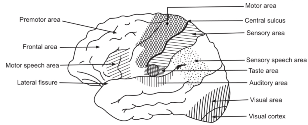
The premotor area lies in the frontal lobe immediately anterior to the motor area. The cells are supposed to exert a controlling influence over the motor area, ensuring orderly movements. In the lower part of this area just about the lateral sulcus, there is a group of nerve cells known as the motor speech area which controls movements necessary for speech. It is dominant in the left hemisphere in right-handed people and vice-versa.
The frontal area extends anteriorly from the premotor area to include the remainder of the frontal lobe. It is a large area. It is more highly developed in human beings than in other animals. It is responsible for the behavior, character, and emotional states of the individual.
Sensory Areas
The post-central (sensory) area lies behind the central sulcus. In this area, sensations of pain, temperature, pressure; touch, knowledge of muscular movement, and the position of joints are perceived. The sensory area of the right hemisphere receives impulses from the left side of the body and vice versa.
The parietal area lies behind the postcentral area and includes the greater part of the parietal lobe of the cerebrum. Its functions are supposed to be associated with obtaining and retaining accurate knowledge of objects. The sensory speech area is situated in the lower part of the parietal lobe and extends into the temporal lobe. The spoken word is perceived here. The auditory (hearing) area lies immediately below the lateral sulcus within the temporal lobe. The cells receive and interpret impulses transmitted from the inner ear by the vestibulocochlear (auditory) nerves. The olfactory (smell) area lies deep within the temporal lobe where impulses from the nose via the olfactory nerves are received and interpreted. The tasting area lies just above the lateral sulcus in the deep layers of the sensory area. In this area, impulses from special nerve endings in taste buds in the tongue and in the lining of the cheeks, palate, and pharynx are perceived as taste. The visual area lies behind the parietal-occipital sulcus and includes the greater part of the occipital lobe. The optic nerves (nerves of the sense of sight) pass from the eye to this area. It receives the impulses as visual impressions.
Other Areas
Deep within the cerebral hemispheres, there are groups of cell bodies termed as nuclei. They act as relay stations where impulses are passed from one neuron to the next in a chain. Following are important masses of grey matter.
- Basal nuclei
- Thalamus
- Hypothalamus
- Basal nuclei: This area lies deep within the cerebral hemispheres. It is supposed to influence skeletal muscle tone. If control is inadequate or absent, movements are jerky, clumsy, and uncoordinated.
- Thalamus: It consists of two masses of nerve cells and fibers situated within the cerebral hemispheres just below the corpus callosum, one on each side of the third ventricle. Sensory input from the skin, viscera, and special sense organs is transmitted to the thalamus before redistribution to the cerebrum.
- Hypothalamus: It is composed of a number of groups of nerve cells. It is situated below and in front of the thalamus, immediately above the pituitary gland. It is linked to the posterior lobe of the pituitary gland by nerve fibers and to the anterior lobe by a complex system of blood vesicles. It controls the output of hormones from both lobes of the gland. It controls the following additional functions.
- Autonomic nervous system.
- Hunger and thirst. Body temperature.
- Emotional reactions: pleasure, fear, etc.
- Sexual behavior.
- Biological clocks or circadian rhythms, e.g. sleeping and waking cycles
Make sure you also check our other amazing Article on: Anatomy Of The Heart