It is a microscope that uses a beam of an accelerated electron as a source of illumination. The wavelength is 100,000 times shorter than visible photon light. It has higher resolving power than the light microscope. Based on the work, it is four different types analytical electron microscopy (AEM), scanning transmission electron microscope (STEM), Scanning electron microscope (SEM), and transmission electron microscope (TEM).
Table of Contents
Principle:
Analytical electron microscopy (AEM) is a type of microscopy for capturing information on the interaction between the incident electrons and the atoms of the specimen. It is a tool for observing nano-scale structures also. It is a transmission electron microscope equipped with analytical functions such as energy dispersive X-ray spectrometry and electron energy loss spectrometry, to enable qualitative and quantitative measurements, elemental distribution mapping, and chemical state analyses in the micro-size observation areas.
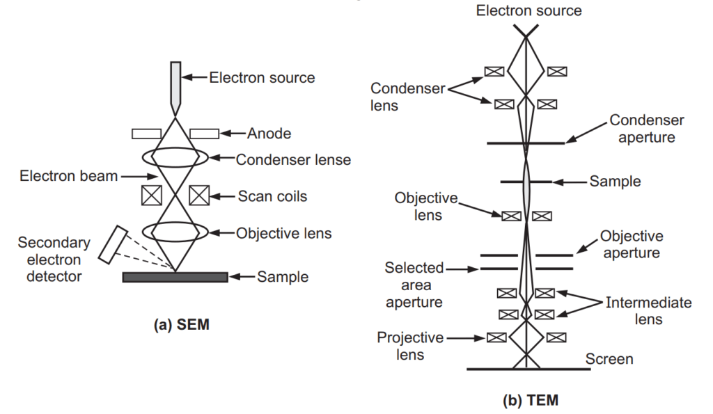
TEM is a microscopy technique in which a beam of electrons is transmitted through an ultra-thin specimen, interacting with the specimen as it passed through it. It is mainly used for cancer research, and virology, to examine small columns of the atom, crystal orientation, etc.
STEM is a modified type of TEM. It uses magnetic lenses to focus a beam of the electron. The image is formed by the primary electrons coming through the specimen.
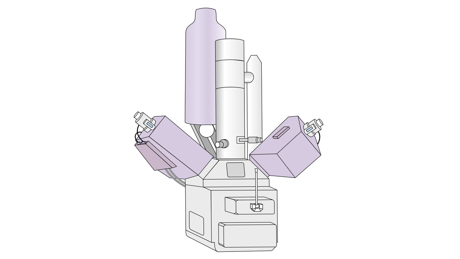
SEM is a microscopy technique that produces images of a sample by scanning it with a focused beam of electrons. The electrons interact with the atoms in the sample and produce various signals that contain information about the sample’s surface topography and composition. It is mainly used in ultra-high vacuum, air, and various liquid states. It is also used for the examination of live specimens (Fig).
Applications:
- The ability to view the microscopic structure of a specimen at a higher resolution.
- It is used for particle analysis or material characterization in a research laboratory.
- It is used to explore the molecular nature and mechanisms of disease.
- It is used to view the 3D structure of biological tissues or cells, determine the structure of proteins and observe viruses in a biological context.
Advantages:
- It is used to study objects of more than 0.2 micrometers.
- It is used for the analysis of subcellular structure.
- It is used for the study of intracellular pathogens and viruses.
- It is used for cell metabolism.
- It is used to study the microstructure of nature.
Disadvantages:
- Instruments are highly expensive.
- The electron microscope is dynamic, which means that the voltage needs to be highly stable.
- The cooling system needs constant circulation pumping through the unit and the vacuum setup also requires consistent pressure and continuous pumping to keep the entire system in proper working condition.
- An electron microscope requires that all samples be viewed in a vacuum. Otherwise, the molecules that occur naturally in the air would scatter and distort the electrons.
Make sure you also check our other amazing Article on : Phase Contrast Microscopy