
Frits Zernike 1953 discovered Phase Contrast Microscopy of a microscope. It is mainly used to visualize unstained living cells. It is an optical microscopy technique, that converts phase shifts in the light passing through a transparent specimen to brightness changes in the image. Phase shifts themselves are invisible, but based on brightness variations, they become visible. This microscope made it possible for biologists to study living cells and their multiplication through cell division.
Principle:
The main principle is based on the small phase changes in the light rays, induced by differences in the thickness and refractive index of the different parts of an object, which can be transformed into differences in light intensity.
In simple terms, it is the translation of invisible phase shifts into visible differences in intensities (Fig). In a phase-contrast microscope, image contrast is increased in two ways: by generating constructive interference between scattered and background light rays in regions of the field, and by reducing the amount of background light that reaches the image plane.
Components:
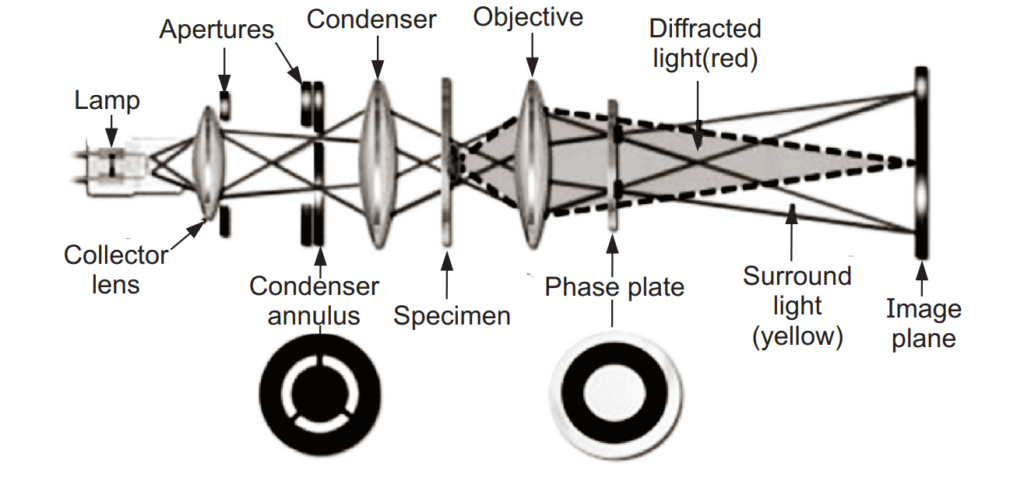
It has a light source, condenser, objective lens system, ocular lens system, annular diaphragm, and phase plate.
Applications:
- It enables the visualization of living and unstained cells.
- It is used for the visualization of various cell organelles like mitochondria, nucleus, vacuoles, etc.
- It helps to study cellular events like cell division, phagocytosis, etc.
- It helps to visualize cellular movements like chromosomal and flagellar movements.
- It is used to observe the growth of the living cells in the plant tissue culture techniques.
- It is used to study the membrane permeability of the cells.
Advantages:
- It provides clear images of unstained cells.
- It provides high-contrast images of the cells.
- It restricts the damage to the cells due to chemical preparation and staining.
- It enhances the prolonged observation of living cells.
- Its cost is affordable.
- It is widely applied in biological and medical research, especially throughout the fields of cytology and histology.
Disadvantages:
- It produces bright holo surrounding the image because of the partial formation of direct and deviated rays.
- It is only effective to observe individual cells.
Make sure you also check our other amazing Article on : Dark Field Microscopy