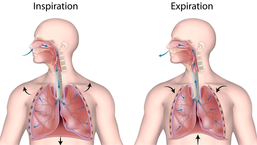
Pulmonary ventilation is the inspiration and expiration of air between the atmosphere and lungs.
Table of Contents
External Respiration
Exchange of gases between the atmosphere and lungs; that is, the blood gains O2 and loses CO2.
Internal Respiration
Internal respiration involves the exchange of gases between blood and tissue cells. The blood gives O2 and receives CO2 from the tissues.
The respiratory system consists of the nose, pharynx (throat), larynx (voice box) trachea (windpipe), bronchi, and the lungs. But structurally, the respiratory system consists of two parts:
- The upper respiratory tract which refers to the nose, pharynx and associated structures.
- The lower respiratory tract refers to the larynx, trachea, bronchi and lungs.
Functionally, there is the conducting part which consists of the nose, pharynx, larynx, trachea, bronchi, bronchioles and terminals of bronchioles. All these conduct air into the lungs. The respiratory part, on the other hand, consists of respiratory bronchioles, alveolar ducts, alveolar sacs and alveoli. This part serves as a site for the exchange of gases.
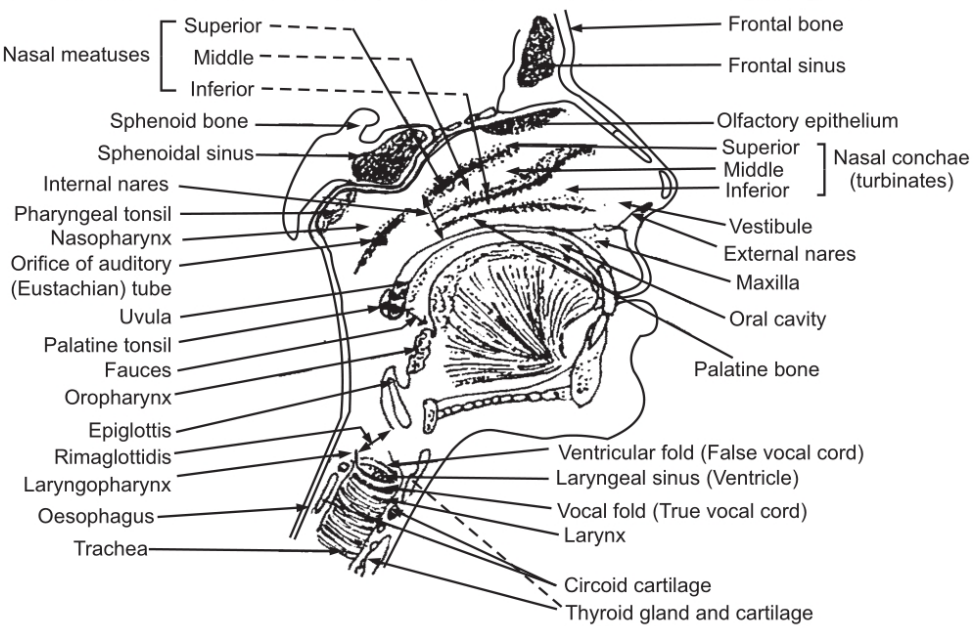
Nose
The nose consists of an external part and an internal part in the side of the skull. The nasal bone forms the bridge of the nose which holds it in a fixed position. It has a framework of pliable hyaline cartilage. The hyaline cartilage is covered with muscle and skin and is lined with mucous membrane. There are two nares or nostrils present on the surface of the external nose. The nose performs three main functions:
- It warm moistens and filter incoming air
- It receives olfactory stimuli.
- Its large hollow resonating chambers modify speech sounds.
Internally, it communicates with the pharynx through two openings called the internal nares. Various sinuses from frontal, sphenoid, maxillary, ethmoidal and lachrymal ducts open into the internal nose; the ethmoidal, maxillae, lachrymal, palatine form walls of the internal nose. The roof is formed by the ethmoidal bone whereas the floor of the nose is formed by palatine bones. The nasal cavity is divided into two parts by the nasal septum. Anteriorly, the septum consists of hyaline cartilage and the remaining part is formed by vomer which is a perpendicular plate of the ethmoidal bone.
From each lateral wall of the nasal cavity, projections arise, which are termed superior, middle and inferior conches. The olfactory receptors lie in the membrane lining the superior nasal conches and adjacent to the septum. This portion is called as the olfactory epithelium. The mucous membrane contains capillaries and ciliated columnar epithelium with many goblet cells. When air enters through the nasal cavity, the blood in the capillaries warms the air while mucous from goblet cells moistens the air and traps dust particles. The cilia move the dust particles towards the pharynx so that they can be eliminated from the respiratory tract by swallowing, spitting, or coughing.
The pharynx is a tube-like structure about 05 inches in length; it lies between internal nares to the cricoid cartilage. It lies superior to the larynx and posterior to the nasal and oral cavity. The walls of the pharynx are composed of skeletal muscles and are lined with mucous membranes. The pharynx provides a passage for food and air and also serves as a resonating chamber for speech sounds. It also shows the presence of tonsils which help to eliminate foreign invaders by immunological reactions.
The nasopharynx lies superior to the pharynx, posterior to the nasal cavity and extends to the level of the soft palate. Overall, there are five openings in the walls of the nasopharynx: two for the internal nares, two for auditory tubes, and one for the oropharynx. For equalization of pressure at the tympanic membrane, the nasopharynx conveys air through auditory tubes. At the posterior wall of the nasopharynx, pharyngeal tonsils are present.
The oropharynx is the intermediate of the pharynx. It lies posterior to the oral cavity and extends from the soft palate and is inferior to the level of the hyoid bone. The portion of the pharynx used for digestive as well as respiratory purposes is called throat or faucets. The palatine and lingual tonsils are present in the oropharynx.
The laryngopharynx starts from the hyoid bone and connects to the oesophagus with the larynx. Both, the oropharynx and laryngopharynx are lined by non-keratinised stratified squamous epithelium.
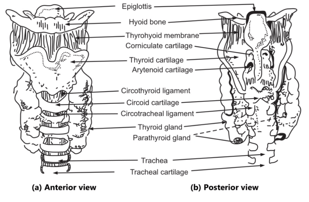
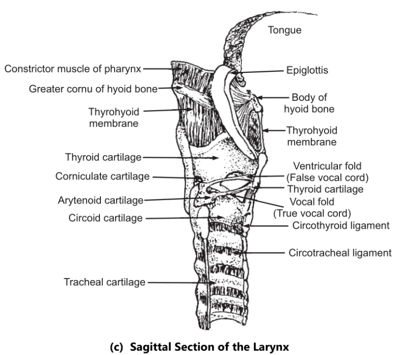
The Larynx or Voicebox
This has a short passage which connects the laryngopharynx with the trachea. It lies between the 4th-6th cervical vertebrae. The larynx is composed of nine pieces of cartilage. The three single cartilages are thyroid cartilage, epiglottis or epiglottis cartilage and cricoid cartilage. The three paired cartilages are arytenoids, cuneiform and articulate cartilages. Arytenoids cartilage influences the position and tension of the vocal cords.
The thyroid cartilage consists of two fused plates of hyaline cartilage which forms the anterior wall of the larynx.
The Epiglottis
It is a large leaf-shaped elastic cartilage piece. The stem is attached to the anterior rim of thyroid cartilage whereas the leaf portion remains unattached. This leaf portion is free to move up and down like a trap door. The opening of the glottis is closed by the epiglottis during swallowing.
The glottis consists of a pair of mucous membrane folds (i.e. vocal cord) in the larynx. When the larynx is closed off, the liquid and food enter into the oesophagus.
The interior wall of the larynx is formed by cricoid cartilage. This is attached to the first ring of the cartilage of the trachea.
Arytenoids Cartilage
These are triangular pieces, composed of hyaline cartilage and are present at the posterior, superior border of the cricoid cartilage. Arytenoids cartilage is attached to vocal folds and intrinsic pharyngeal muscles. Due to the contraction of intrinsic pharyngeal muscles, the movement of the vocal fold takes place. The paired corniculate cartilages support vocal folds and the lateral side of the epiglottis.
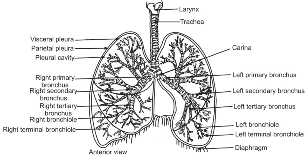
Trachea
It is a tubular passageway for air. It starts from the larynx and extends up to the 5th thoracic vertebra where it divides into the right and left branches. The trachea is approximately 12 cm in length and 2 1/2 cm in diameter. The trachea is composed of 16-20 incomplete ‘C’ shaped rings of hyaline cartilage. These rings are united by fibrous and muscular tissues. These cartilages form the anterior and lateral walls of tubes. The posterior surface of the trachea is flat because of a deficiency of cartilage. This posterior part is composed of fibrous and elastic tissues with involuntary muscle fibres. These rings are enclosed by fibrous and elastic tissues. The trachea exhibits three coverings. The outer surface is composed of fibrous and elastic tissues, which encloses rings of hyaline cartilage. The middle layer is composed of fibrous and elastic tissue lined with areolar connective tissue. Through this passes the lymphatic and blood vessels. The inner layer is lined by ciliated epithelial cells which chiefly contain goblet cells responsible for the secretion of mucous. The blood is supplied by inferior thyroid arteries; venous blood is collected by inferior thyroid veins. The nerve is supplied by the vagus and recurrent laryngeal nerve.
Bronchi
The bronchi commence at the fifth thoracic vertebra. The right bronchus is a shorter and wider tube and assumes a more vertical position. It is 02.5 cm in length. The right bronchus immediately after entering the right lung, gives off a branch. The branches continue to divide in the lung tissues. The left bronchus is narrower and longer. It is 05 cm long. The left bronchus too, after entering into the left lung, divides into many branches. The bronchi also contain incomplete rings of hyaline cartilage. These are lined by pseudostratified ciliated columnar epithelium.
The Bronchioles
The bronchi divide into smaller and smaller branches called bronchioles. These bronchioles do not have cartilage in their walls. Initially, they are composed of muscle tissue, fibrous tissue and elastic tissues with an inner lining of cubical epithelium. As the tubes become smaller, fibrous and muscular tissues disappear and the cubical epithelium is converted into a single layer of flattened epithelium. The minute branches of bronchioles are known as terminals of bronchioles. These further bifurcate to form respiratory bronchioles. The respiratory bronchiole is expanded at its to form an alveolar duct. The alveolar duct leads to minute sac-like structures called alveoli, where the exchange of gases occurs between the air in the alveoli and blood in the capillaries.
Make sure you also check our other amazing Article on: Creatinine Phosphate