Anatomy of the Ear, the Ear is the origin of hearing. The VIII cranial nerve carries the sensation of hearing towards the cerebral cortex of the brain in the hearing area where interpretation occurs.
Most part of the ear is situated in the petrous part of the temporal bone except the pinna lying outside.
Table of Contents
Structure of the Ear
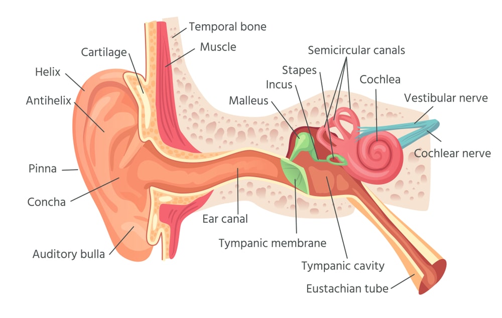
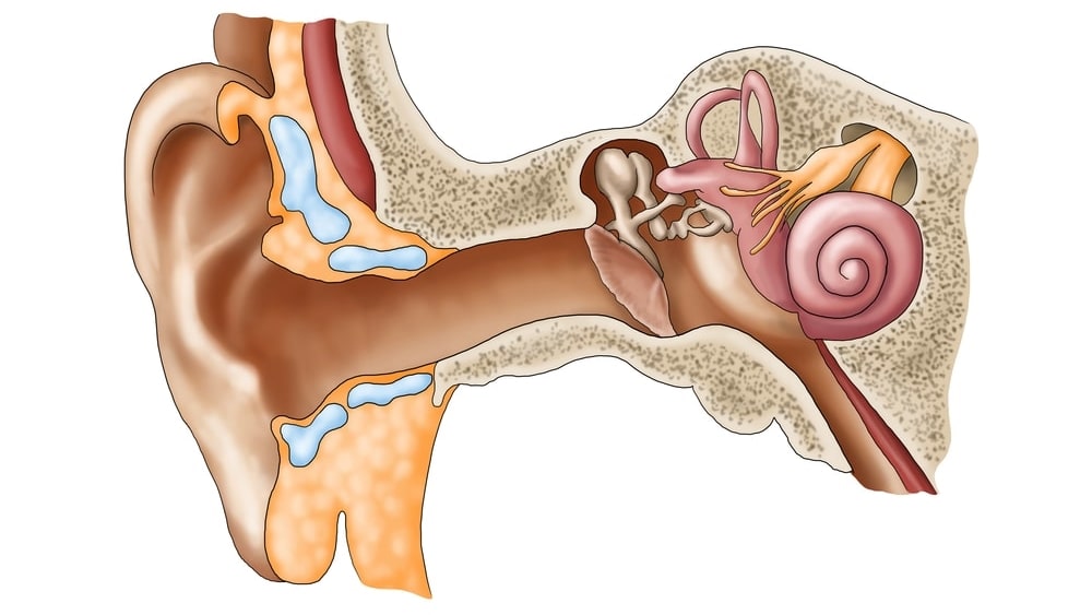
The ear has three parts: external ear, middle ear, and internal ear.
External Ear
The external ear consists of the pinna and external auditory meatus.
Pinna or auricle is the outer part of the ear which is an expanded part, projecting from the side of the head. This part is made of fibroelastic cartilage which is covered with the skin. The size of the pinna is variable in different animals. The expanded outer edge is known as the helix and the lowermost part is known as the lobule. It is composed of fibrous and adipose tissue, supplied with blood capillaries.
External Auditory Meatus
It is an irregular-shaped long tube about 02.5 cm in length; it starts from the pinna to reach the tympanic membrane or eardrum. It is divided into two parts: The cartilaginous part which is about one-third of the canal and the osseous part, lying medially two-third of the canal. The ceremonious glands lie in the cartilaginous portion. The ceremonious glands are modified sweat glands which secrete wax called ceriman. The ceriman helps to trap the unwanted particles entering in the ear, such as dust or insects.
The tympanic membrane or eardrum separates the external ear from the middle ear. The eardrum is round and oval in shape and composed of three layers of tissues. The outer layer is made up of stratified epithelium; the middle layer is made up of fibrous tissue, and the inner layer contains cuboidal epithelium.
The Middle Ear
It is also known as a tympanic cavity. It is the irregular-shaped cavity lying in the temporal bone. It is filled up with air by the auditory tube which comes from the pharynx. The auditory tube is about 4 cm long. Air reaches the cavity through the Eustachian tube which extends from the nasopharynx. The pressure on the eardrum is maintained by air coming from the atmosphere. through the external auditory meatus and air coming through the auditory tube. So the eardrum remains in a stretched condition which helps for vibration due to sound waves. The lateral side of the middle ear is made up of the tympanic membrane, roof, and the floor is formed by temporal bone. The posterior wall is also made up of temporal bone. It has a small opening which extends to the mastoid atrium. It is used for passing air to reach mastoid cells.
In the medial wall, there are two openings, an oval window or fenestrate vestibule where a small bone called stapes fits and the round window or fenestrate cochleae which are closed by a thin sheet of fibrous tissue.
The middle ear contains the chain of small three bones called auditory ossicles. One end of the auditory ossicle is attached to the eardrum and the other to the fenestrate vestibule. The auditory ossicle contains small bones called, the malleus, the incus, and the stapes. The malleus is hammer-shaped which contains a head, neck, and handle. The handle of the malleus is attached to the eardrum and the head shows a movable joint with incus. The incus is anvil-shaped, present in the middle part of a chain. It contains a head, neck, two limbs, and a base. The body is attached to the malleus and process with the stapes. The stapes are stirrup-shaped; contain a head, neck, two limbs, and a base. The body is attached to the malleus and process with the stapes. The stapes are stirrup-shaped; contain a head, neck, two limbs, and a base. The head of stapes is fixed with incus and the base is fixed in the fenestrate vestibule. With the help of ligaments, these three bones maintain their position.
The Internal Ear
It contains a bony portion, called the bony labyrinth, and a membranous portion called the membranous labyrinth. The cavity of the bony labyrinth is larger than the membranous labyrinth. The membranous labyrinth fits into the bony labyrinth. There is a space between two labyrinths that contains a fluid called per lymph whereas membranous labyrinth also contains a fluid called endolymph. The bony labyrinth contains three parts: the vestibule, the cochlea, and three semicircular canals. The expanded part near the middle ear is known as a vestibule, containing the vestibule and fenestra cochlea. The cochlea looks like a snail’s shape which has a broad base and the apex. The semi-circular canals are continuous with the vestibule. The shape of the membranous labyrinth is similar. This part is smaller than a bony labyrinth. The vestibule contains utricle and saccade. The membranous cochlea has the duct of the cochlea. The inside part is known as a basilar membrane. On the basilar membrane lies the nerve cells and nerve fibers. The cells are long and narrow and situated side by side and hair is present on the cells. All nerve cells and nerve fibers form a true organ of hearing called the organ of a corgi. The nerve fiber combines with each other to form the auditory nerve-VIII cranial nerve. The auditory nerve goes backward through the temporal bone to reach the hearing area of the cerebral cortex of the brain.
Physiology of Hearing
Any sound in the atmosphere produces the sound waves or vibrations and the sound waves travel at a certain speed, say about 1089 feet/per meter/second. Because of the shape of the auricle, it captures any sound wave and relays it through the auditory canal, so that eardrum can vibrate. Due to the vibration of the eardrum, the auditory ossicle moves to and fro and it sets the per lymph in motion and end lymph present inside the membranous labyrinth gets stimulated, and thus kept in motion.
The nerve cells present on the basilar membrane get stimulated; and consequently, the nerve fibers carry impulse through the auditory nerve to the hearing area of the cerebral cortex, where the interpretation of the waves take place.
Disorder of Ear
Deafness: There are many reasons for deafness, it may due to improper sound transmission in the external ear or damage to the neural pathways. The nerve deafness may be caused by acoustic nerve degeneration or tumors in the acoustic meatus. Whereas conducting deafness may be caused by wax deposition in the external auditory meatus or damage to auditory ossicles or thickening of the eardrum etc.
Make sure you also check our other amazing Article on: Anatomy Of Eye