It is a type of differential staining where two or more stains are used to differentiate between types of bacteria. The gram staining technique was discovered by Hans Christian Gram in the year 1884.
Importance of the Gram Staining
- It is an important test for the rapid diagnosis of infectious agents.
- It differentiates types of bacteria whether Gram-positive or Gram-negative.
- It helps in the study of the morphology of bacteria.
- It helps in finding the pieces of evidence of capsule, spores, pus cells, epithelial cells, Yeast cells, etc.
- To understand how the Gram stain reaction affects Gram-positive and Gram-negative bacteria based on the biochemical and structural differences of their cell walls.
Steps follow on the gram staining
- Crystal violet: It is the primary stain that is used as a simple stain because it dyes the cell wall of bacteria cells.
- Gram’s Iodine: It acts as a mordant which helps to fix the primary dye to the cell wall.
- Decolourizer: It is used to remove the stain of primary dye (crystal violet) from the Gram-negative bacterial cell wall. Decolourizer is composed of organic solvents like acetone, ethanol, or in combinations.
- Safranin: Finally, a counterstain is applied to stain those cells that have lost their primary stain (especially Gram-negative bacteria) as a result of decolorization.
The procedure is based on the ability of microorganisms to retain the color of the stains used during the gram stain reaction. Gram-negative bacteria are decolorized by the alcohol, losing the color of the primary stain, purple. Gram-positive bacteria are not decolorized by alcohol and will remain purple.
After the decolorization step, a counterstain is used to impart a pink color to the decolorized gram-negative organisms.
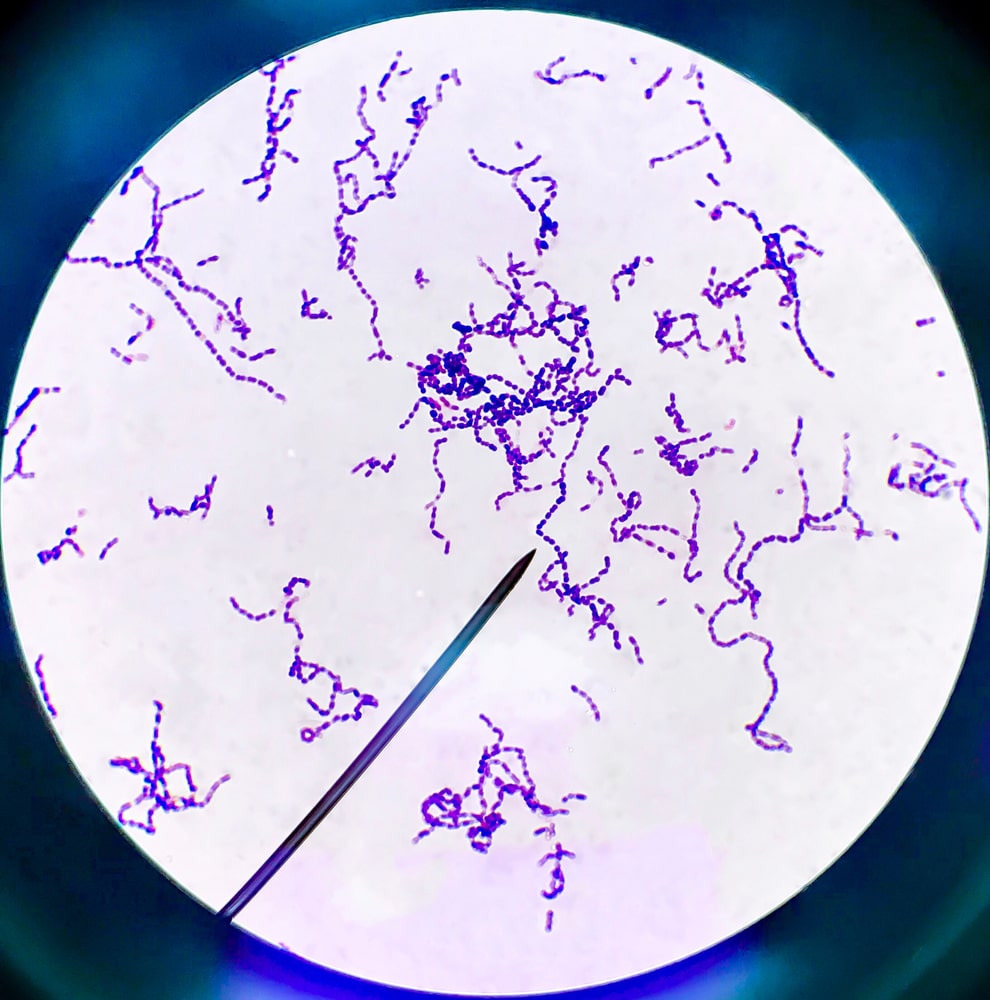
Gram-positive bacteria: Stain dark purple due to retaining the primary dye called Crystal violet in the cell wall. Example: Staphylococcus aureus.
Gram-negative bacteria: Stain red or pink due to retaining the counterstaining dye called Safranin.
Example: Escherichia coli.
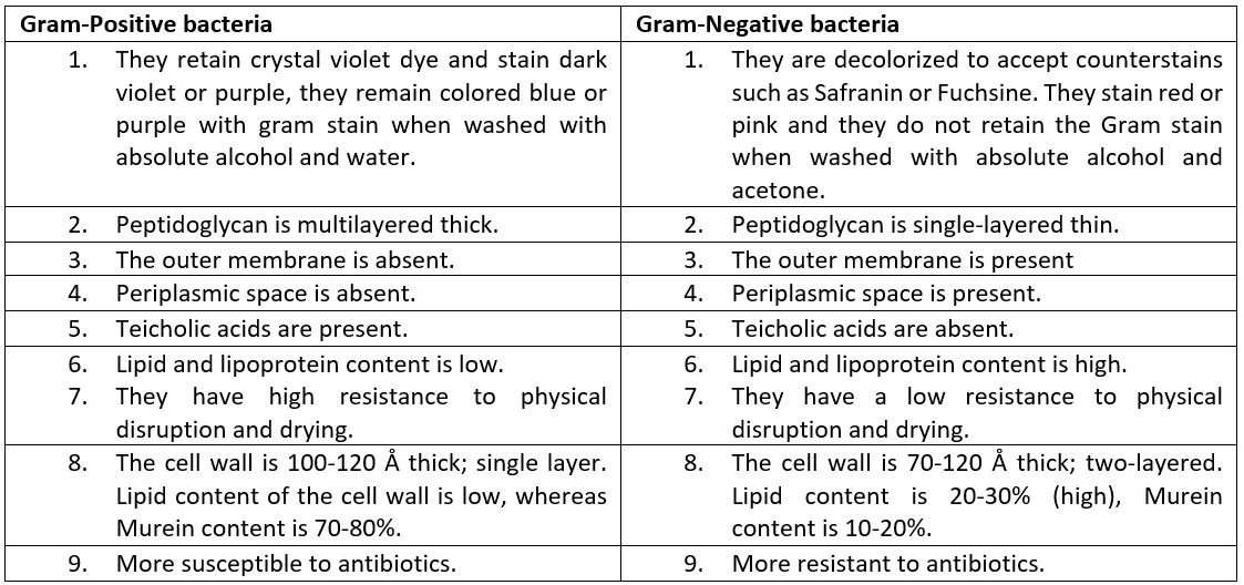
There are some differences between Gram-positive and Gram-negative bacteria which are listed in Table 1.1
Table 1.1 Differences between Gram-Positive and Gram-negative bacteria
Make sure you also check our other amazing Article on : Simple Staining