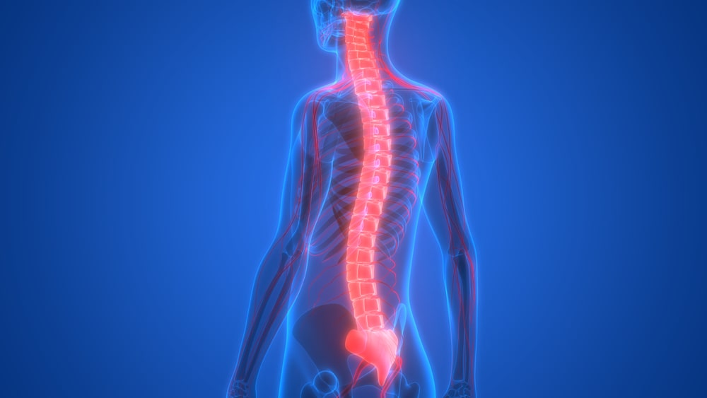
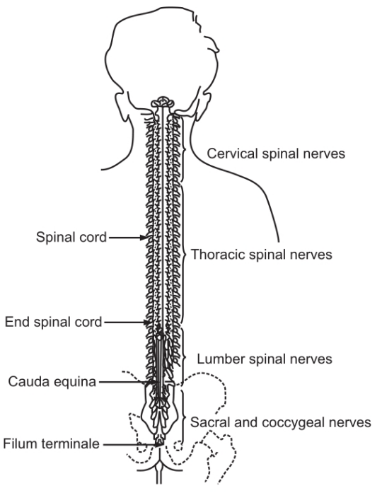
It is the elongated and almost cylindrical part extending from the brain just below the medulla oblongata. It is suspended in the vertebral canal and is surrounded by meninges and cerebrospinal fluid. It extends from the first cervical vertebra to the lower border of the first lumbar vertebra. It is approximately 45 cm long and about the thickness of the little finger. Except for the cranial nerves, the spinal cord is the nervous tissue link between the brain and the rest of the body. Motor nerves originating from the brain descend through the spinal cord and supply to various organs and tissues at appropriate levels of the cord. Sensory nerves from different organs and tissues enter and pass upwards to the brain via the spinal cord. Some activities like spinal reflexes are independent of the brain. In such cases, motor action is decided and implemented at the level of the spinal cord itself. In order to facilitate spinal reflexes, there are extensive neuron connections between sensory and motor neurons at the same or different levels in the spinal cord.
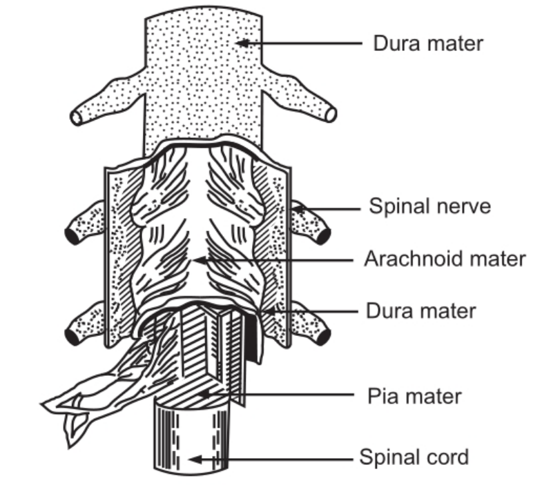
Each cut away to show the underlying layers
A cross-section of the spinal cord shows that it is composed of grey matter in the center and is surrounded by white matter supported by neuralgia.
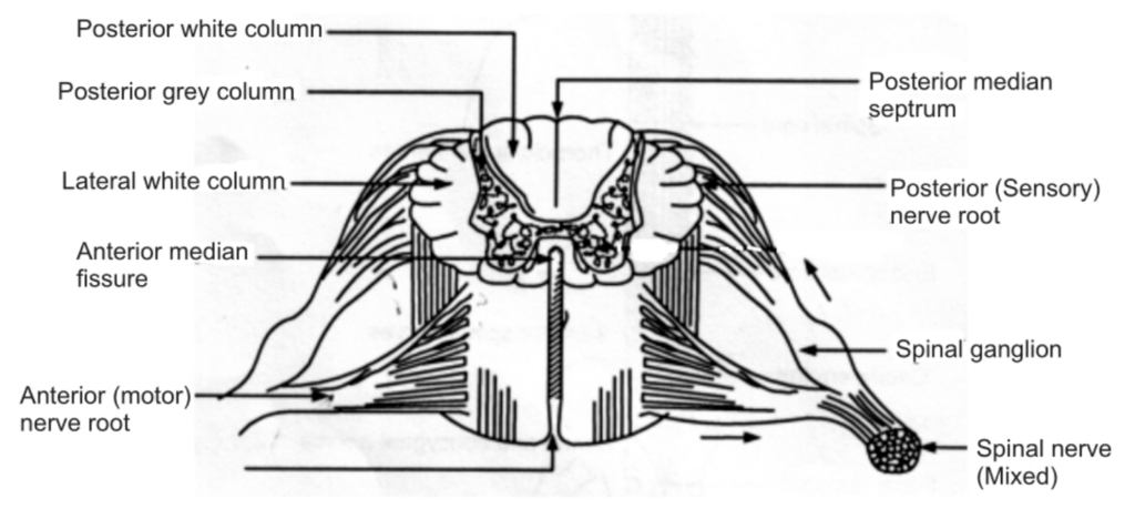
The arrangement of grey matter in the spinal cord resembles the shape of the letter H, having two anterior, two posterior, and two lateral columns. The area of the grey matter lying transversely is the transverse commission and is pierced by the central canal. The canal extends from the fourth ventricle in the brain and contains cerebrospinal fluid. The spinal cord consists of the following cell bodies:
Sensory cells receive impulses from the periphery of the body. Lower motor neurons transmit impulses to the skeletal muscles. Spinal nerve (Mixed) Connector neurons, linking sensory and motor neurons at the same or different levels.
Posterior columns of grey matter are composed of cell bodies which are stimulated by sensory impulses from the body. The nerve fibers of these cells form white matter and transmit the sensory impulses to the brain.
Anterior columns of grey matter are composed of the cell bodies of the lower motor neurons; they are stimulated by the axons of the upper motor neurons or by the cell bodies of connector neurons. All sensory nerve fibers pass through the posterior root ganglia. These ganglia promote the onward movement of nerve impulses from the periphery.
The white matter of the spinal cord is arranged in three columns or tracts; anterior-posterior and lateral. These tracts are formed by sensory nerve fibers ascending to the brain, motor nerve fibers descending from the brain, and fibers of connector neurons.
Table of Contents
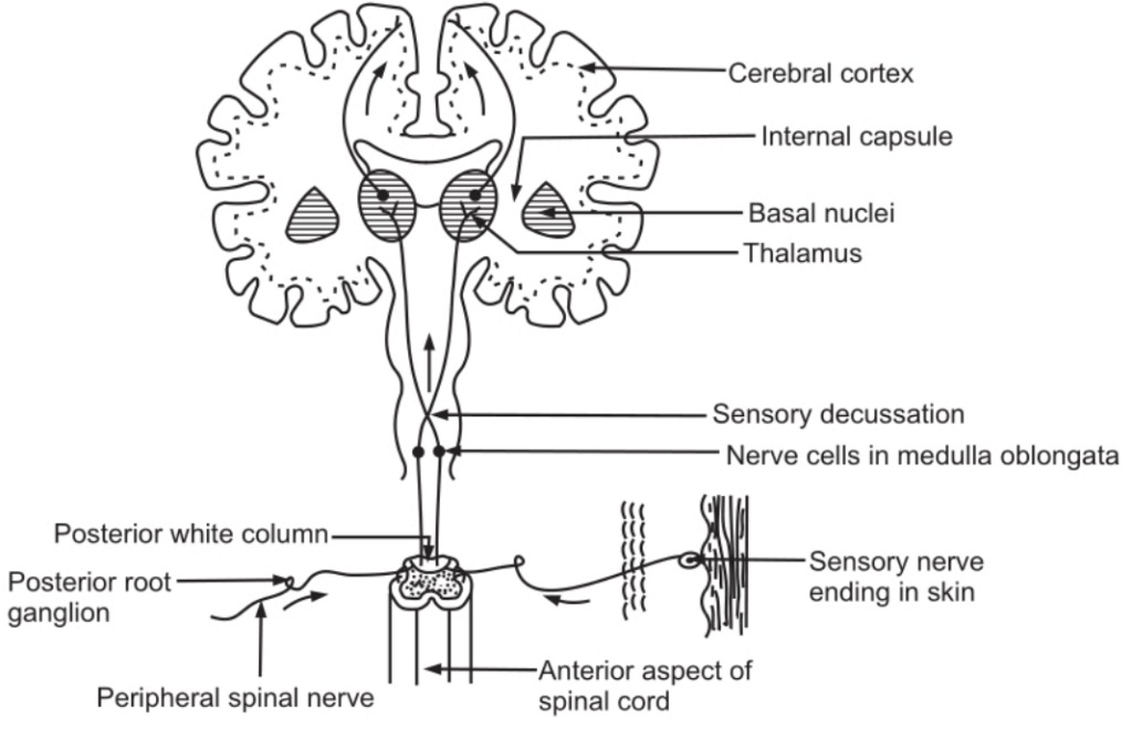
Sensory Nerve Tracts in the Spinal Cord
Following are two main sources of sensation transmitted to the brain via the spinal cord:
- The Skin: Sensory nerve endings in the skin are called as cutaneous receptors. They are stimulated by pain, heat, cold; touch, and pressure. The sensory impulses are passed to the opposite hemisphere of the cerebrum and the sensation is perceived in that region.
- The tendons, muscles, and joints: Sensory nerve endings in these structures are called proprioceptors and are stimulated by the stretch. Along with impulses from the eyes and ears, they are associated with maintenance of balance and posture and with the perception of the position of the body in space.
Motor Nerve Tracts in the Spinal Cord
Neurons which transmit nerve impulses away from the brain are termed as motor neurons. insulation of neurons results in the following actions:
- Contraction of skeletal muscles.
- Contraction of smooth muscles and the secretion by glands controlled by nerves of the autonomic nervous system.
Movements of Voluntary Muscles
The contraction of the muscles moving the joints is a voluntary act. The stimulus for this act originates at the level of consciousness in the cerebrum. Some impulses are originated in the midbrain, brainstem, and cerebellum. This activity occurs below the level of consciousness and is associated with coordination of muscle activity.
Efferent nerve impulses are transmitted from the brain via bundles of nerve fibers for tracts through the spinal cord. There are two types of tracts in the spinal cord:
- Pyramidal (corticospinal)
- Extra pyramidal
The motor fibers forming pyramidal tracts travel through the internal capsule and are the main pathway for impulses to skeletal muscles. Those motor fibers which do not pass through the internal capsule form the extra pyramidal tracts and have connections with many parts of the brain including the basal nuclei and the thalamus.
The upper motor neuron has its cell (Betz’s cell) in the precentral sulcus of the cerebrum. The axons pass through the internal capsule, pons, and medulla. In this spinal cord, they form the lateral corticospinal tracts and these fibers terminate in close association with the cells of lower motor neurons in the anterior columns of grey matter. The axons of these upper motor neurons make up the pyramidal tracts and decussate in the medulla oblongata, forming the pyramids.
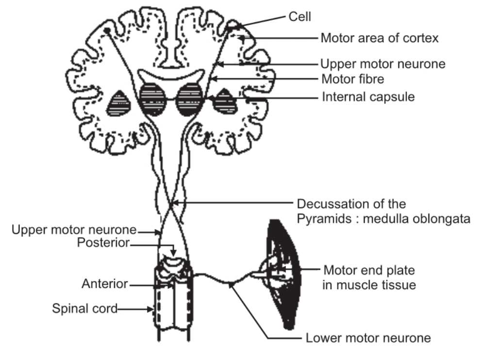
The lower motor neuron has its cell in the anterior horn of grey matter in the spinal cord. Its axon emerges from the spinal cord by the anterior root, joins with the incoming sensory fibers, and forms the mixed spinal nerve. At its termination in muscle the axon branches into a variable number of tiny fibers which form motor end-plates. Each motor end plate has a close association with a sensitive area on the wall of a muscle fiber. The motor end-plates of each nerve fiber and the muscle fibre form a motor unit.
At the end of the motor unit, acetylcholine is secreted after stimulation of the nerve. Acetylcholine is termed as a neurotransmitter Secretion of this neurotransmitter is responsible for the contraction of smooth or skeletal muscles.
The lower motor neuron is the final common pathway for the transmission of nerve impulses to skeletal muscles. The cell of this neuron is influenced by various upper motor neurons. Some of these neurons stimulate the cells while others inhibit it. A balance between these two actions results in smooth, coordinated muscle movement. A part of this movement may be voluntary and a part of it may be involuntary in nature.
Movements of Involuntary Muscles
Upper motor neurons have their cells in the brain at a level below the cerebrum that is in the midbrain, brainstem, and cerebellum or spinal cord. They influence muscle activity in relation to the maintenance of posture and balance, the coordination of muscle movement, and the control of muscle tone.
Spinal reflexes consist of three elements:
- Sensory neurons.
- Connector neurons.
- Lower motor neurons.
In the reflex arc, only one of the elements exists. A reflex action is an immediate motor response to a sensory stimulus. Many connectors and motor neurons may be stimulated by afferent impulses from a small area of the skin. These, in turn, stimulate many connectors and lower motor neurons in the cord which results in the contraction of many skeletal muscles of different organs. Reflex action takes place very quickly. It is of a protective type; they may be inhibited occasionally.
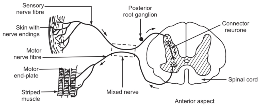
In stretch reflexes, only two neurons are involved. The cell of the lower motor neuron is stimulated by the sensory neuron. There is no connector neuron involved in it, e.g. knee jerk.
By tapping the tendon just below the knee when it is bent, the sensory nerve endings in the tendon, and in the thigh muscles are stretched. This initiates a nerve impulse which passes into the spinal cord to the cell of the lower motor neuron in the anterior column of grey matter on the same side. As a result, the thigh muscles suddenly contract, and the foot kicks forward.
Make sure you also check our other amazing Article on: Brain Stem