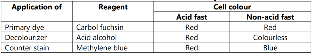
The acid-fast staining method is one of the differential staining techniques. This technique was first developed by Ziehl and later on modified by Neelsen and hence this method is also known as Ziehl-Neelsen staining techniques. In the year 1883, Neelsen used Ziehl’s carbol-fuchsin and heated then decolorized it with acid alcohol followed by counterstained with methylene blue. Thus Ziehl-Neelsen staining technique was developed as acid-fast staining (Table 1.1).
Objectives:
- To differentiate bacteria into an acid-fast group and non-acid fast groups.
- It is used for those microorganisms which are not staining by simple or Gram staining method, particularly the member of genus Mycobacterium which is resistant to other stains.
Principle of the Acid Fast Staining
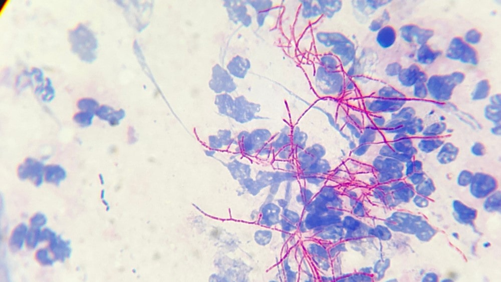
The smear is prepared and stained with carbol fuchsin which solubilizes the lipoidal material present in the Mycobacterial cell wall and by heating, carbol fuchsin further penetrates through the lipoidal wall and enters into the cytoplasm, resulting in all cells appearing red. Then the smear is decolorized with 3% HCl in 95% alcohol (decolourizing agent) but the acid-fast cells are resistant due to the presence of a large amount of lipoidal material in their cell wall that prevents the penetration of the decolourizing solution. Then the smear is stained with a counterstain, methylene blue, and only decolorized cells are absorbed and appear blue while acid-fast cells retain the red color (Table 1.1 ).
The procedure involved in the Acid Fast Staining
- Prepare bacterial smear on the clean and grease-free slide, using sterile technique.
- Allow smear to air dry and then heat fix.
- Cover the smear with carbol fuchsin stain.
- Heat the stain until vapor just begins to rise about 60°C. Allow the heated stain to remain on the slide for 5 minutes.
- Wash off the stain with clean water.
- Cover the smear with 3% v/v acid alcohol for 5 minutes or until the smear is sufficiently decolorized, i.e. pale pink.
- Wash well with clean water.
- Cover the smear with malachite green stain for 1–2 minutes, using the longer time when the smear is thin.
- Wash off the stain with clean water.
- Wipe the back of the slide clean, and place it in a draining rack for the smear to air[1]dry.
- Examine the smear microscopically, using the 100 X oil immersion objective.
Example of Acid-fast
Mycobacterium tuberculosis, Mycobacterium smegmatis.
Non-Mycobacterial bacteria: Nocardia; Coccidian Parasites: Cryptosporidium.
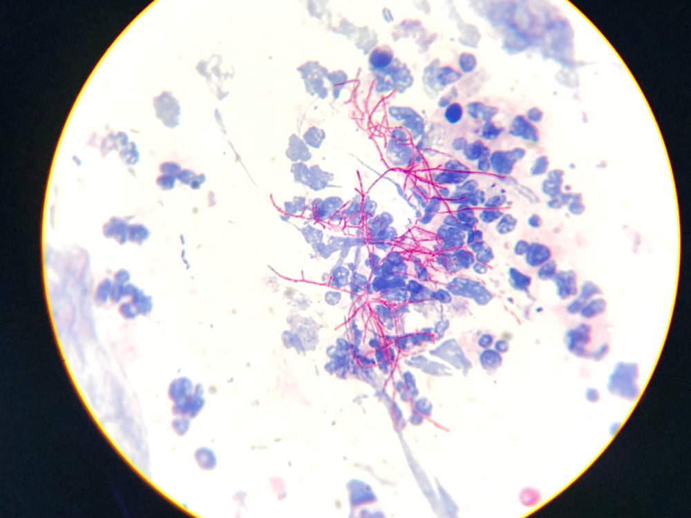
slightly curved rods
Make sure you also check our other amazing Article on : Gram Staining